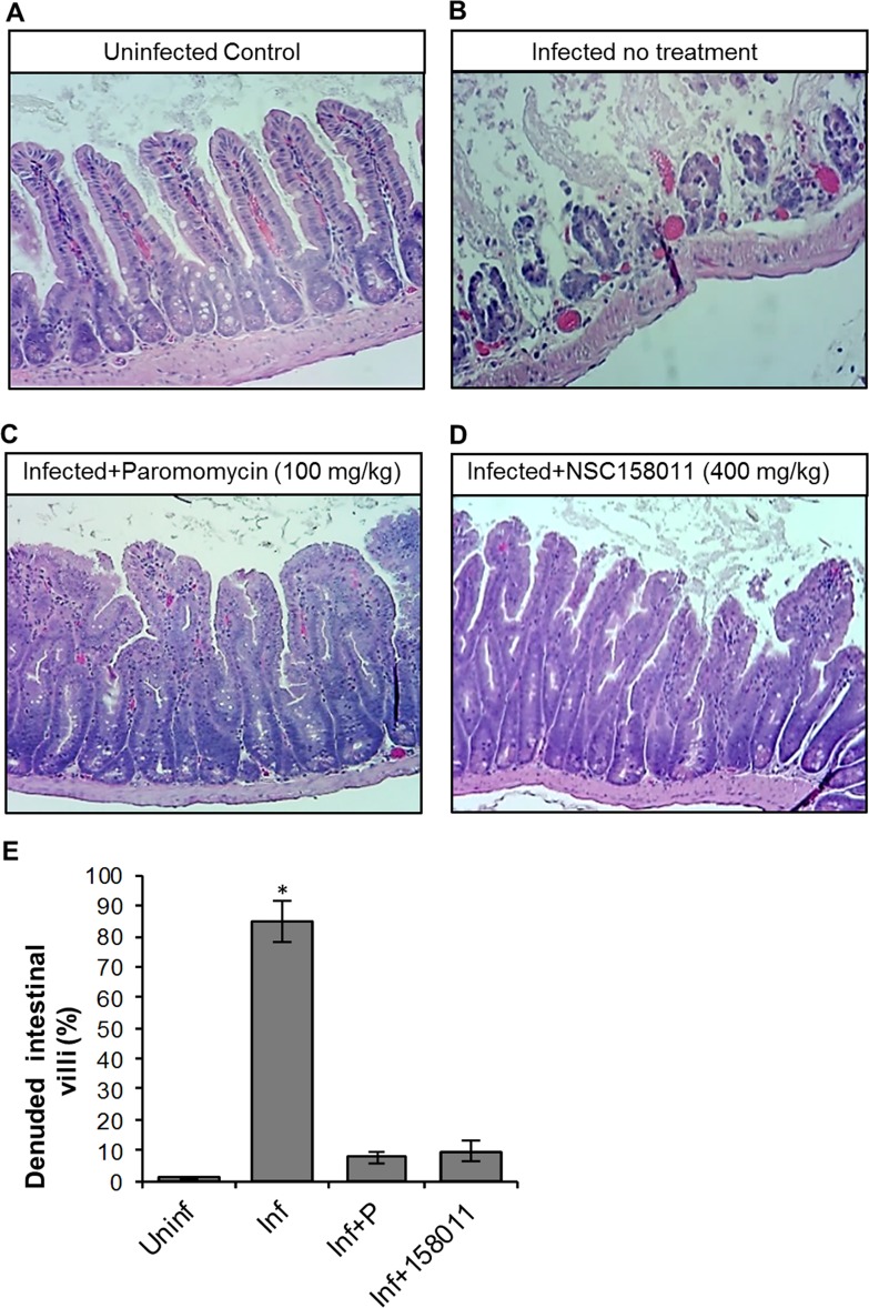
Fig 7. Histopathological analysis of the effect of Cryptosporidium parvum infection in the lower small intestines of mice with or without NSC158011 treatment.
Mice infected with C. parvum were maintained untreated (Infected no treatment), treated with paromocycin (Infected+Paromomycin (100 mg/kg)) or treated with compound NSC158011 at 400 mg/kg (Infected+NSC158011 (400 mg/kg)) for 8 days. Uninfected mice were maintained as control (Uninfected Control). After 8 days, mice were sacrificed and the lower intestinal tissue processed for histology and stained with hematoxylin and eosin. (A) Uninfected control mice samples depicted intact intestinal epithelium with prominent villi. (B) In contrast, infected mice without treatment depicted denuded villi. Both (C) paromomycin and (D) NSC158011 treated infected mice depicted intact intestinal epithelium with prominent villi that were comparable to the uninfected control. The images are representative of samples analyzed from 3 mice per treatment group. (E) The mean percentage of denuded intestinal villi in 4 randomly chosen microscopic fields per sample from the uninfected (Uninf), infected untreated (Inf), Infected treated with paromomycin (Inf+P), and infected treated with 400 mg/kg NSC150811 (Inf+150811) mice. The data shown represent means for samples from three mice per group with standard error bars and levels of statistical significance depicted (*P < 0.05).

