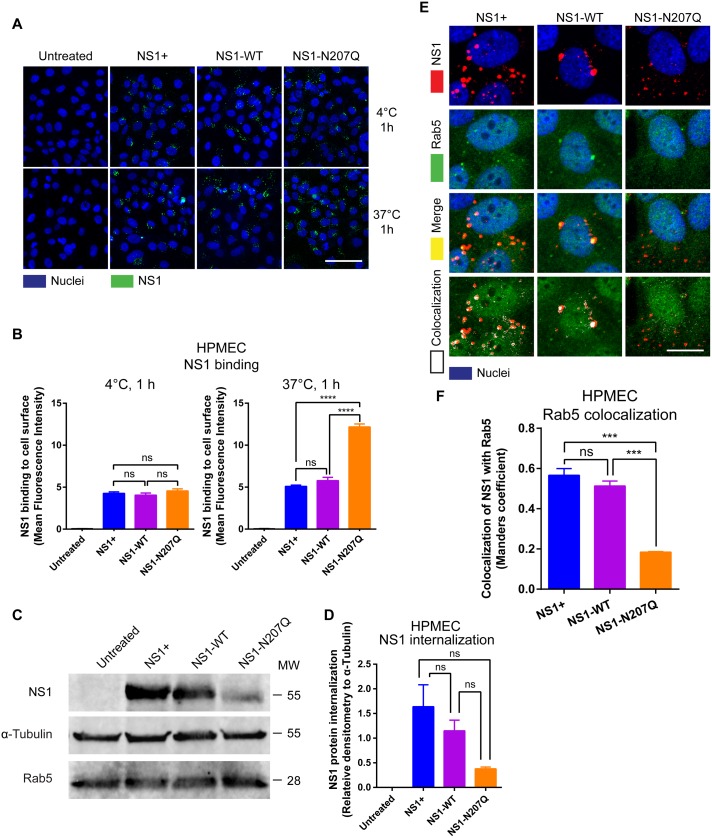
Fig 3. Mutation of the N-glycosylation site 207 of DENV NS1 results in defective NS1 internalization.
(A) NS1 protein binding (green) to HPMEC 1 hpt at 4°C (top row) or 37°C (bottom row) was visualized via IFA. Images (20X; scale bars, 50 μm) are representative of 2 independent experiments. (B) MFI was used to quantify the amount of NS1 binding to the cell surface 1 hpt at 4°C and 37°C in Fig 3A. The means ± SEM of two individual experiments run in duplicate are shown. ns = not significant; ****, p<0.0001. (C) Confluent HPMEC monolayers were exposed to 10 μg/ml of different NS1 proteins and incubated at 37°C for 1 hours. Trypsin was used to remove surface-bound NS1, and cell lysates were analyzed by Western blot. Western blot shows detection of the internalized DENV NS1 protein, GAPDH (loading control), and Rab5, an early endosome marker. (D) Quantitation of C. Each bar represents the mean ± standard error of the mean (SEM) of densitometry values normalized to the control protein α-tubulin from two independent experiments. (E) Co-localization of NS1 proteins (red), as indicated, with Rab5 (green) in HPMEC. Co-localization is shown in yellow in merge image or white in co-localization panel (JACoP, ImageJ). Nuclei are stained with Hoechst (blue). Images (40X; scale bars, 10 μm) are representative of 2 individual experiments run in duplicate. NS1+, NS1 from Native Antigen Company. (F) Quantification of DENV NS1 and Rab5 colocalization in HPMEC in Fig 3E. Quantification of the amount of spatial overlap between the two signals (NS1 in red and Rab5 in green) in Fig 3E was obtained using four different frames from the maximum projections of two RGB images based on the object-based approach (JACoP) and defined by the Manders’ Coefficient as previously described [59]. The colocalization coefficient was normalized taking into account the signal obtained from 300 cells per image. Each bar represents the mean ± SEM of MFI values in the colocalization analyses obtained from two independent experiments. NS1+, NS1 from Native Antigen Company. ns = not significant; ***, p<0.001.

