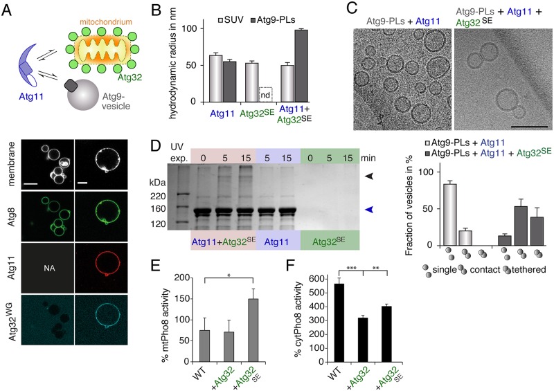
Fig 5. The cargo receptor Atg32 activates Atg11.
(A) Atg8Atto488, enzymatically coupled to Atto633-labeled GUVs, fails to recruit PacificBlue-labeled Atg32, in which the AIM is mutated (Atg32WG). Recombinant Atg11Atto565 recruits Atg32WG to Atg8-decorated GUVs. Scale bar 10 μm. (B) The RH of SUVs (dark grey bars) and Atg9-PLs (light grey bars) as obtained in DLS experiments in the presence of either Atg11 or Atg32SE or of both. RH of polydisperse samples is not determinable (nd). (C) Cryo-EM of mixtures of Atg9 vesicles with Atg11 or with Atg11 and Atg32SE as indicated. The chart shows the number of single, contact, and tethered vesicles as fraction of all vesicles of the indicated sample. At least 40 vesicles were analyzed in each of three pools. One pool represents data from different EM grids of identical samples. Single vesicles do not contact any other vesicle; contact vesicles are in close proximity to other vesicles with an apparent contact between membranes. Tethered vesicles have a flattened and extended contact area as shown in the corresponding image. Scale bar 100 nm. (D) SDS-PAGE of samples from cross-linking experiment using the UV-activatable reagent Sulfo-LC-SDA with a spacer arm of 12.5 Å. Sulfo-LC-SDA was conjugated to primary amines in Atg32SE. Atg11, Sulfo-LC-Atg32SE or mixtures of both were exposed to 365 nm UV light for indicated times. The blue arrowhead indicates the band corresponding to Atg11 and the black arrowhead that of the crosslinked species with an apparent molecular weight of approximately 350 kDa. (E and F) Mitochondrial Pho8ΔN60 assay (E, mtPho8) of log-phase growing and cytoplasmic Pho8ΔN60 assay (F, cytPho8) of starved WT cells and cells overexpressing Atg32 or Atg32SE lacking its transmembrane domain. Pho8 activity was corrected for total protein amount of each sample and normalized to the signal of WT cells (100%). (B) Data are presented as mean values ± SD of n = 5 measurements. (E and F) Mean values ± SD of n = 3 independent experiments are shown. P values were calculated using a two-tailed Student t test (*P < 0.05, **P < 0.01). See also S4 Fig. AIM, Atg8-interacting motif; DLS, dynamic light scattering; EM, electron microscopy; GUV, giant unilamellar vesicle; PL, proteoliposome; RH, hydrodynamic radius; SUV, small unilamellar vesicle; WT, wild-type.

