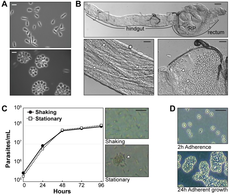
Fig 1. Crithidia fasciculata adherent stages in culture and the mosquito host.
(A) Phase contrast images of live swimming (top panel) and adherent rosettes (bottom panel) grown in culture. Scale bars, 10 μm. (B) Images of Crithidia rosettes within an infected Aedes aegypti hindgut. Top panel shows an infected hindgut. C. fasciculata colonize the hindgut, rectum and rectal papillae (RP). Scale bar, 100 μm. Bottom panels are enlargements of the areas shown in the boxes above. Scale bars, 20 μm. Adherent cells are visible throughout the highlighted regions. White arrowheads in bottom panels indicate rosettes (C) Growth curve comparing rates of growth of cells grown in flasks kept on a rocker (shaking, solid line) and the incubator shelf (stationary, dotted line). Images of cells in shaking and stationary flasks. White arrowhead indicates a rosette. Scale bar, 25 μm. (D) Top image, cells that were allowed to adhere for 2 h followed by 3 washes to remove swimming cells. Bottom image shows the same flask after adherent cells have been allowed to grow for ~24 h. Scale bar, 25 μm.

