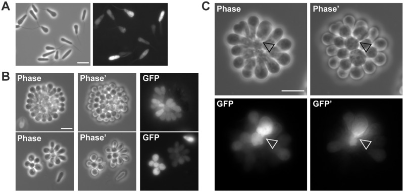
Fig 2. Organization of cultured C. fasciculata rosettes.
(A) Live C. fasciculata swimming cells expressing cytoplasmic GFP from an episomal plasmid. Scale bar, 10 μm. (B) Live GFP-expressing rosettes grown on poly-L-lysine-coated glass coverslips. Two different focal planes are shown in phase contrast to highlight the three-dimensional structure of the rosette. Scale bar, 10 μm. (C) Enlargement of rosette cells expressing GFP. Arrowhead indicates possible junction between cells. Scale bar, 10 μm.

