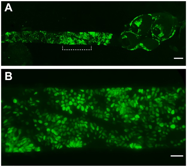
Fig 4. Hindgut of Ae. aegypti mosquito 7 days post infection with GFP-expressing C. fasciculata.
(A) Widefield fluorescence microscopy was used to image fluorescent parasites in a dissected mosquito hindgut. Parasites are observed attached to the hindgut, rectum and rectal papillae. (B) Higher magnification image of the bracketed region indicated in panel A. The scale bars in panels A and B are 100 μm and 20 μm, respectively.

