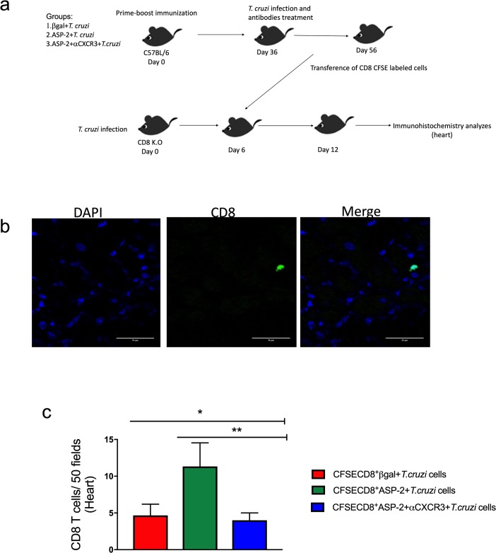
Fig 7. The treatment with anti-CXCR3 decreases the number of CD8+ T cells in the heart.
a-Experimental design showing the immunization, infection, treatment with anti-CXCR3 antibody and adoptive transference of CD8+ CFSE labeled cells to CD8 deficient mice (infected). Briefly, C57BL/6 mice were immunized with prime-boost heterologous protocol, the first dose of immunization was with pCDNA3/pIgSPclone9 after 21 days of prime the mice received Adβgal/AdSP-2 and after 15 days were infected with T. cruzi and treated with 250 μg of CXCR3 antibody, and on day 20 after infection, CD8+ T cells from spleen were purified, labeled with CFSE, and transferred into CD8 K.O mice previously infected (on day 6 after infection). After 6 days of transference, the number of CD8+ T cells in heart from βgal+T.cruzi, ASP2+T.cruzi and ASP2+αCXCR3+T.cruzi groups was quantified. b-IHC in heart showing the CFSE+CD8+ T cells (green). The DAPI staining was used to label the nucleus of the cells. c-Number CFSE+CD8+ T cells that migrated to CD8 K.O mice hearts. The cells were counted using the fluorescent microscopy and 50 fields were counted. Results are shown as individual values and the mean ± SEM for each group (n = 3). Statistical analysis was performed using the One-Way ANOVA and Tukey’s HSD tests. Symbols indicate that the values observed were significantly different between the groups (*p = 0.010; **p < .05).

