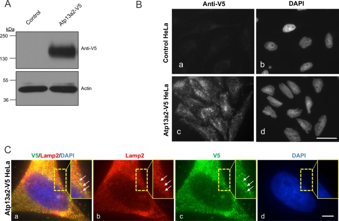
Fig 1. Isolation of a cell line overexpressing human Atp13a2.
(A) HeLa cells stably expressing Atp13a2-V5 or the parental control HeLa cells were lysed and equal amounts of protein were probed with a V5 antibody to detect expression of the V5-tagged Atp13a2 protein by immunoblotting. Actin was used to verify protein loading. (B) Control HeLa cells (a and b) or HeLa cells stably expressing Atp13a2-V5 (c and d) were grown on coverslips and stained with the anti-V5 antibody followed by a FITC-conjugated secondary antibody and examined by immunofluorescence microscopy. The anti-V5 antibody decorated puncta in the stable cell line, which is consistent with localization of the Atp13a2-V5Isoform-1 protein to lysosomes. (C) HeLa cells stably expressing Atp13a2-V5 were double stained with anti-V5 and anti-Lamp 2 antibodies and their binding was detected with Alexa 488-conjugated and Alexa 594-conjugated secondary antibodies. The images shown are of the same cell imaged for the two different fluorescent proteins and DAPI, which was used to stain the nucleus. The result of merging of the three individual channels is also shown. The stippled box is a magnified portion of the cell to better illustrate the colocalization of V5 and Lamp 2 staining (white arrows). Bar, 10 μm.

