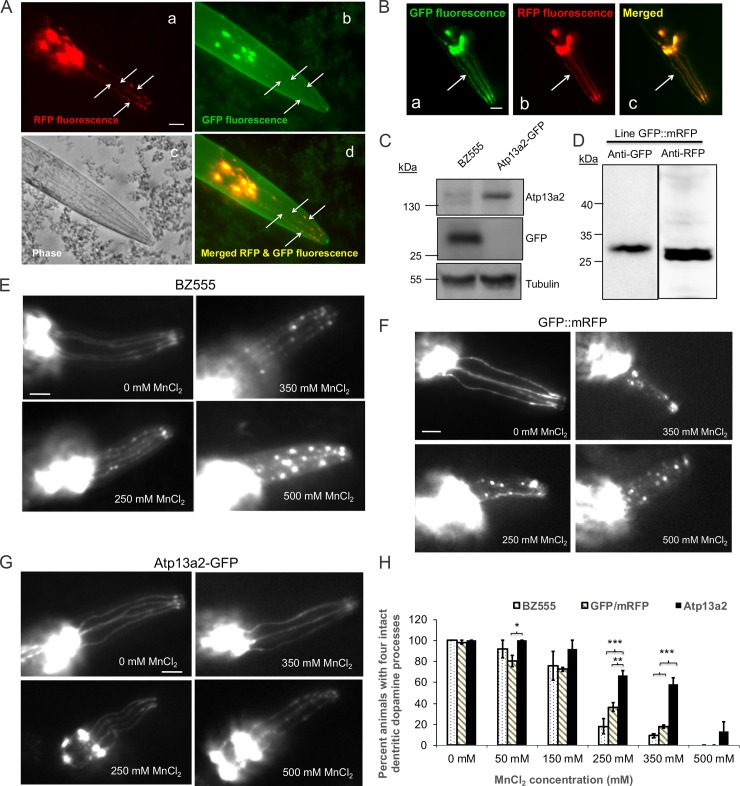
Fig 5. Nematodes expressing GFP-tagged Atp13a2 exhibit higher resistance to manganese induced dopamine neuron degeneration.
(A) Double fluorescence microscopy demonstrating visual detection of RFP and GFP fluorescence in dopamine neuron processes (indicated with arrows) in the head of the C. elegans line coexpressing dat-1p:mRFP and dat-1p:Atp13a2-GFP. The phase contrast image of the head region is also shown, together with the image produced after merging the red and green fluorescent images. Because of its brighter fluorescence the RFP fluorescence was used to monitor dopamine neuron degeneration after manganese exposure. Bar, 25 μm. (B) Fluorescent images of the head region of the nematode line coexpressing dat-1 promoter driven expression of GFP (a) and mRFP (b) and the result of merging the two fluorescent proteins (c). The arrow shows clear colocalization of GFP and mRFP fluorescence in dopamine neurons. (C) Immunoblots of C. elegans lysates prepared from the BZ555 and Atp13a2-GFP lines with antibodies specific for Atp13a2, GFP or tubulin. The immunoblots confirmed expression of the appropriate size Atp13a2-GFP protein and GFP proteins in the two different lines. (D) Immunoblot of the GFP::mRFP coexpressing line for GFP and RFP expression. (E, F and G) Representative images showing differential sensitivity of dopamine neurons in the nematode BZ555 line (E) GFP and mRFP expressing nematodes (F) and the Atp13a2-GFP-expressing nematode line (G) to acute exposure for 1 h with different concentrations of MnCl2. Bar, 25 μm. (H) Quantification of dopamine neuron degeneration in the BZ555, GFP::mRFP and Atp13a2-GFP expressing nematode lines. The quantification shown is the mean ±SDM of three independent experiments. At least 20 nematodes were counted for each exposure condition. Nematodes expressing Atp13a2-GFP exhibit significantly higher resistance to manganese-induced neurodegeneration at 250 and 350 mM MnCl2 treatment (*p < 0.05, **p < 0.01, ***p < 0.001).

