
Figure 1.
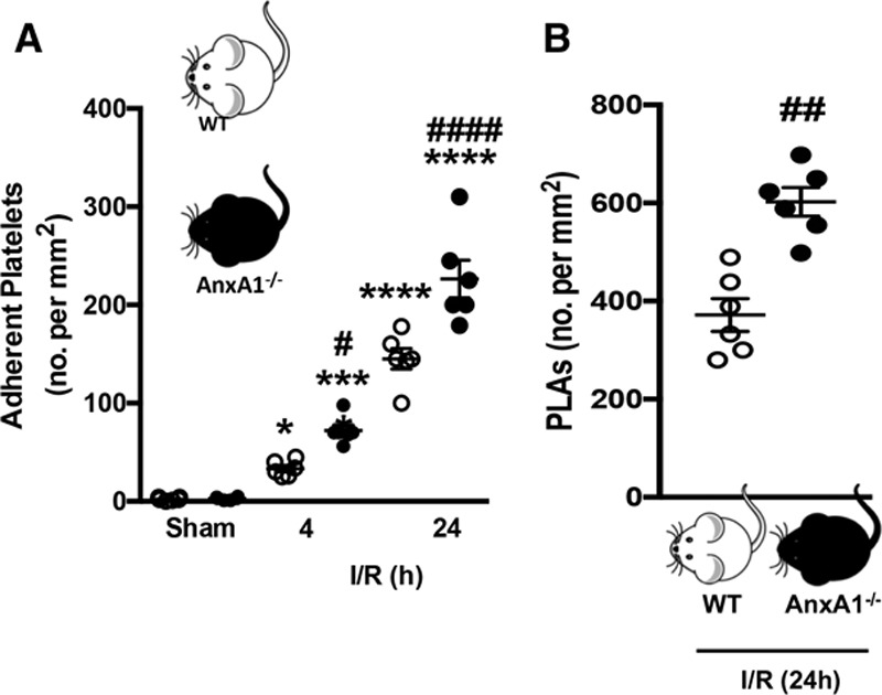
Platelet and platelet–leukocyte interactions in the cerebral microcirculation are heightened in annexin A1 knockout (AnxA1−/−) mice after ischemia reperfusion injury (I/R). Wild-type (WT) and AnxA1−/− mice were subjected to transient middle cerebral artery occlusion for 60 min, followed by 4- or 24-h reperfusion. Intravital fluorescence microscopy was performed to assess cellular interactions in the cerebral microcirculation (pial vessels) of mice subjected to cerebral I/R. Platelets were labeled with carboxyfluorescein succinimidyl ester (90 µmol/L), and leukocytes were labeled with rhodamine 6G (0.02%). Platelet interactions were quantified in terms of numbers (no.) of adherent platelets on the endothelium (A, cells stationary for ≥2 s) and platelets interacting directly with adherent leukocytes on the endothelium (B), termed platelet–leukocyte aggregates (PLAs). The data are means±SEM of 6 mice per group with 2 to 3 vessels per mouse and assessed by ANOVA with Bonferroni post hoc test (A) or Mann–Whitney test (B). *P<0.05, ***P<0.001, and ****P<0.0001 vs sham control of the same genotype. #P<0.05, ##P<0.01, and ####P<0.0001 vs a different genotype at the same time point.