Figure 3.

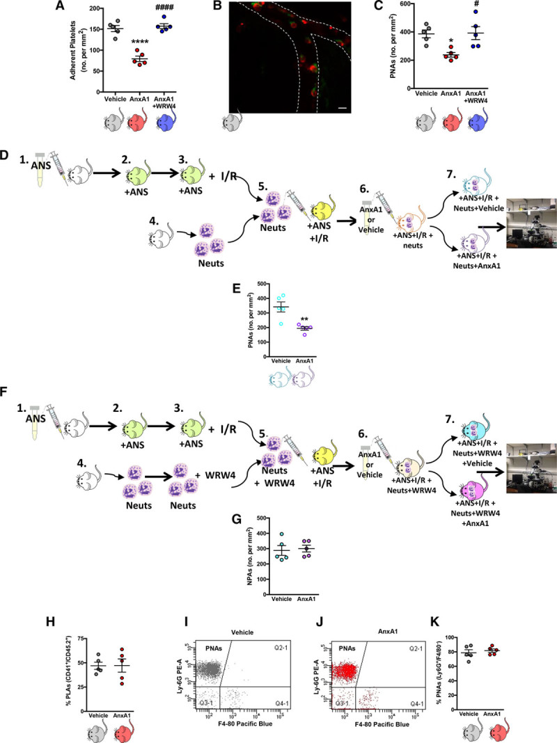
Administration of exogenous annexin A1 (AnxA1) moderates platelet interactions in the brain microcirculation after ischemia reperfusion injury (I/R). Wild-type mice were subjected to transient middle cerebral artery occlusion for 60 min, followed by 24 h of reperfusion (tMCAo/R). A, Vehicle, AnxA1 (3.3 mg/kg), or WRW4 (1.8 mg/kg) were administered intravenously at the start of reperfusion and numbers (no.) of platelet adhesion (cells stationary for at least 2 s) were quantified. B, Representative image of cerebral platelet-neutrophil aggregates (PNAs) 24 h after reperfusion as measured by confocal intravital microscopy. Neutrophils (Neuts, arrow) were labeled with eFluor 488 (green)-labeled anti-mouse Ly-6G, 2 μg/mouse. Platelets (arrowhead) were labeled with Dylight 649 (red)-labeled antimouse CD42, 1 μg/mouse. The scale bar indicates 20 μm. C, Quantification of endothelial-PNAs in the cerebral microcirculation. D, Adoptive transfer experiments were performed by injecting neutrophils isolated from donor mice into neutropenic recipient mice. In steps 1 and 2, recipient mice were rendered neutropenic by administration of antineutrophil serum (ANS). In step 3, neutropenic mice were subjected to cerebral I/R by tMCAo/R. In step 4, neutrophils were isolated from donor mice. In step 5, neutrophils were injected into neutropenic recipient mice subjected MCAo/R. In step 6, the mice were then treated with vehicle (saline) or AnxA1 (3.3 mg/kg) for 30 min before intravital microscopy, which is shown in step 7. E, Quantification of PNAs in the cerebral microcirculation from D using intravital microscopy. F, Adoptive transfer experiments were performed by injecting neutrophils isolated from donor mice into neutropenic recipient mice. In steps 1 and 2, recipient mice were rendered neutropenic by administration of ANS. In step 3, neutropenic mice were subjected to cerebral I/R by tMCAo/R. In step 6, the mice were then treated with vehicle (saline) or AnxA1 (3.3 mg/kg) for 30 min before intravital microscopy, which is shown in step 7. G, Quantification of NPAs (neutrophil platelet aggregates) in the cerebral microcirculation from F using intravital microscopy. H through K, Blood samples were taken from wild-type mice subjected to MCAo, followed by a 24-h reperfusion and treatment with vehicle (saline) or AnxA1 and analyzed by flow cytometry to assess systemic aggregate formation: quantification of circulating. H, Platelet–leukocyte aggregates (PLAs, CD45.2+, and CD41+). I and J, Flow cytometry population groups of PNAs, which are denoted within the CD45.2+, CD41+ population as Ly-6G+, and F4/80− aggregates. K, Quantification of circulating PNAs. The data are means±SEM of 5 mice/group and assessed by ANOVA with a Bonferroni post hoc test (A and B), Mann–Whitney test (E), or a Student t test (G, H, and K). *P<0.05 and ****P<0.0001 vs vehicle (saline)–treated control. #P<0.05 and ####P<0.0001 vs AnxA1.
