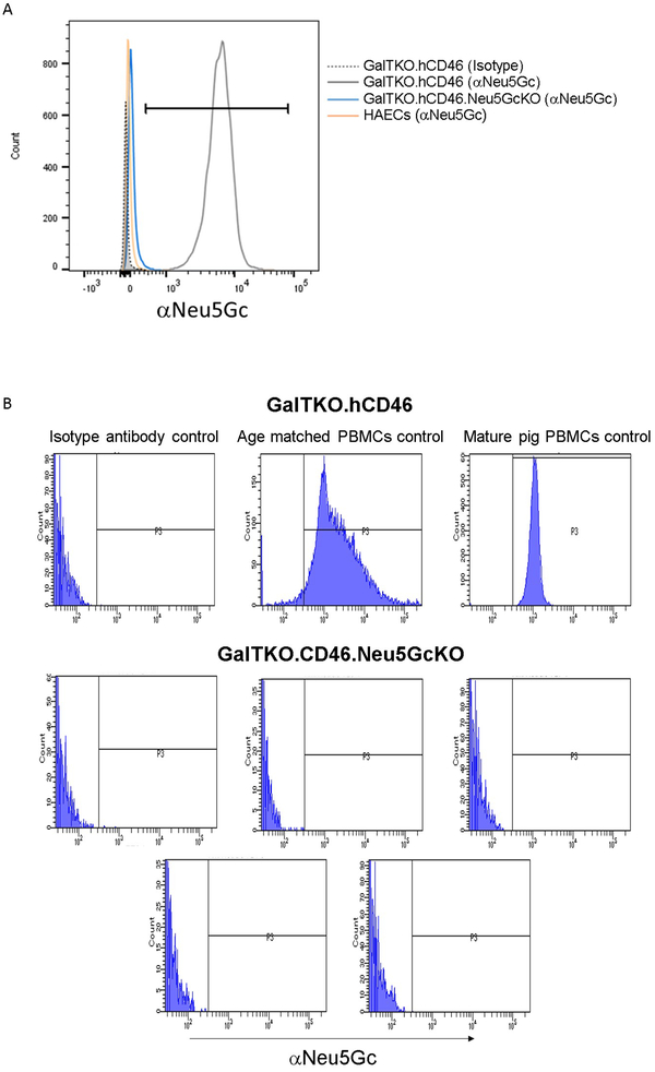
Figure 2: Neu5Gc expression.
Expression of Neu5Gc was assessed on human and pig cells by flow cytometry. A. Human aortic endothelial cells (orange) stained minimally for Neu5Gc (likely from culture media containing fetal bovine serum. Cultured porcine aortic endothelial cells from GalTKO.hCD46 pigs (gray) stained strongly for Neu5GCc compared minimal staining in GalTKO.hCD46.Neu5GcKO endothelial cells (blue). B. Pattern of Neu5Gc expression on peripheral blood mononuclear cells (PBMCs) from GalTKO.hCD46 pigs as a function of pig age; absence of Neu5GcKO expression in PBMCs from GalTKO.hCD46.Neu5GcKO pigs.

