
Fig. 4.
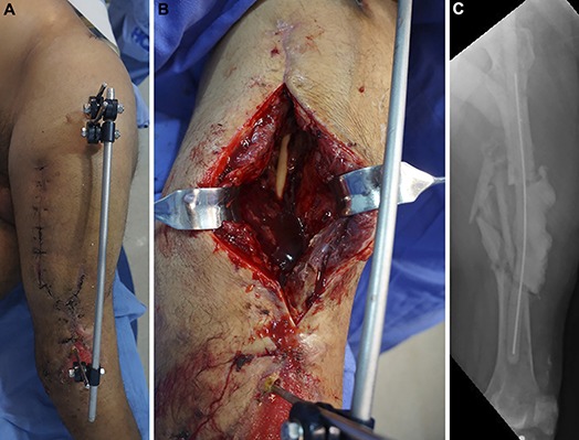
Day 58 after the injury. The spacer had been in the bone defect for 27 days. The patient underwent another procedure to remove the external fixator, obtain tissue samples for culture, and change the spacer in an attempt to prepare for internal fixation. Fig. 4-A Preoperative appearance of the soft tissue. Fig. 4-B Intraoperative photograph showing no clinical signs of infection. Fig. 4-C Radiograph made after removal of the external fixator and insertion of a new intramedullary PMMA-with-antibiotic spacer, which was larger than the previous one to provide stability.