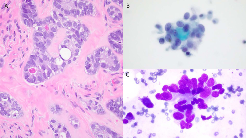
Figure 3.

Cases 3 and 4. Histology shows a low-grade adenocarcinoma with tubular architecture composed of cells exhibiting relatively uniform nuclei with small nucleoli, and occasional hyaline globules (A). Cytology: Mildly pleomorphic small cells with scant cytoplasm, oval nuclei and nuclear molding (B and C), and hyaline globules (B).
