
Figure 3. Activating PAG-USV neurons is sufficient to elicit USV production in the absence of social cues.
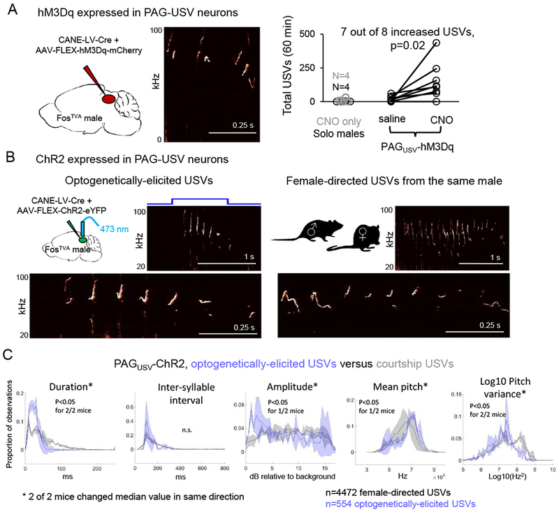
(A) (Left) Schematic of the viruses injected into the caudolateral PAG of a FosTVA male to express hM3Dq in PAG-USV neurons. (Middle) Spectrogram showing USVs produced by a CNO-treated PAGUSV-hM3Dq male. (Right) Total USVs produced in a 60 min. solo test period are shown for different groups of males in chemogenetic experiments: control males (no virus, no CNO, black), males not expressing hM3Dq but treated with CNO (gray), and males with CANE-driven expression of hM3Dq in PAG-USV neurons that were treated with saline (i.p.) or CNO (p = 0.02 for PAGUSV-hM3Dq mice saline versus CNO USVs, Wilcoxon signed-rank test). (B) Optogenetic activation of PAG-USV neurons elicits USVs from males in the absence of female cues. Spectrograms comparing optogenetically-elicited USVs (left) and female-directed USVs (right) from the same male. Bottom panels show expanded views. (C) Distributions of 5 acoustic parameters are shown for optogenetically-elicited USVs (blue) and female-directed USVs (gray) for 2 PAGUSV-ChR2 mice. Asterisks indicate acoustic parameters whose median value changed significantly in the same direction for both mice (p < 0.05, Mann Whitney U tests). See also Figs. S3–5.