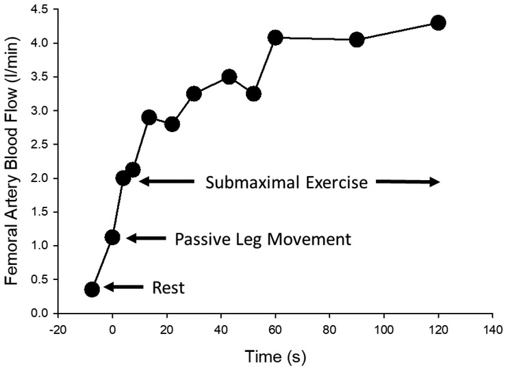
Figure 2: First evidence of peripheral hemodynamic response to passive exercise/movement.
Femoral artery blood flow (ml/min) at rest, during passive leg exercise/movement, and during submaximal single leg knee-extension exercise. Blood flow was measured by Doppler ultrasound during the transition from baseline (no movement) to 60 rpm, which was accomplished within 5 to 7 passive leg movements. Although not the focus of this investigation, this is the first evidence of increased leg blood flow during passive leg exercise/movement. Modified from Radegran and Saltin 1999 [46].

