Abstract
Background:
The association between cerebrospinal fluid (CSF) leaks at the skull base and raised intracranial pressure (ICP) has been reported since the 1960s. It has been suggested that spontaneous CSF leaks might represent a variant of idiopathic intracranial hypertension (IIH). We review the evidence regarding the association between spontaneous CSF leaks and IIH, and the role of ICP in the pathophysiology of nontraumatic skull base defects. We also discuss the management of ICP in the setting of CSF leaks and IIH.
Evidence Acquisition:
References were identified by searches of PubMed from 1955 to September 2018 with the terms “idiopathic intracranial hypertension” and “cerebrospinal fluid leak.” Additional references were identified using the terms “pseudotumor cerebri,” “intracranial hyper-tension,” “benign intracranial hypertension,” and by hand search of relevant articles.
Results:
A CSF leak entails the egress of CSF from the subarachnoid spaces of the skull base into the surrounding cavitary structures. Striking overlaps exist regarding demographic, clinical, and radiological characteristics between IIH patients and those with spontaneous CSF leaks, suggesting that some (if not most) of these patients have IIH. However, determining whether a patient with spontaneous CSF leak may have IIH may be difficult, as signs and symptoms of raised ICP may be obviated by the leak. The pathophysiology is unknown but might stem from progressive erosion of the thin bone of the skull base by persistent pulsatile high CSF pressure. Currently, there is no consensus regarding the management of ICP after spontaneous CSF leak repair when IIH is suspected.
Conclusions:
IIH is becoming more widely recognized as a cause of spontaneous CSF leaks, but the causal relationship remains poorly characterized. Systematic evaluation and follow-up of patients with spontaneous CSF leaks by neuro-ophthalmologists will help clarify the relation between IIH and spontaneous CSF leaks.
The association between cerebrospinal fluid (CSF) leaks at the skull base and raised intracranial pressure (ICP) has been reported for more than 5 decades in the otorhinolaryngology and neurosurgery literature. In the 1960s, Om-maya et al (1) suggested that nontraumatic CSF leaks should be categorized into those related to high ICP or normal ICP. In the 1990s, otorhinolaryngologists (2) emphasized that spontaneous CSF leaks might represent a subset of high-pressure CSF leaks. Therefore, spontaneous CSF leaks might be a presentation of idiopathic intracranial hypertension (IIH). In these patients, the leak acts as a “natural” CSF diversion, and these patients usually do not develop the typical symptoms and signs of intracranial hypertension. Surprisingly, although IIH is routinely diagnosed and managed by neuro-ophthalmologists, very few studies regarding CSF leaks in IIH patients have been published in the ophthalmology or neurology literature (3,4). This highlights the need for IIH specialists to become more knowledgeable regarding spontaneous CSF leaks.
We review the evidence regarding the association between spontaneous CSF leaks, IIH, and ICP in the pathophysiology of nontraumatic skull base defects. We also discuss the management of ICP in the setting of CSF leaks and IIH.
DEFINITION AND CLASSIFICATION OF CEREBROSPINAL FLUID LEAKS
A CSF leak entails the egress of CSF from the subarachnoid spaces of the anterior or middle skull base into the surrounding sinonasal or middle ear cavities through a dehiscence of the lamina dura. Although most patients with CSF leaks at the level of the spine have symptoms of intracranial hypotension (5,6), those with CSF leaks at the level of the anterior or middle skull base often present with isolated clear fluid leakage (rhinorrhea or otorrhea). Spinal CSF leaks have only rarely been associated with intracranial hypertension (7) and will not be further discussed. In this article, the term “CSF leak” will refer to CSF leaks at the skull base unless otherwise specified.
CSF leaks have been variously classified in the literature, but the general principles of a categorization based on mechanisms as developed by Ommaya et al (1) are widely accepted. CSF leaks can be traumatic (80%) or nontraumatic (20%), the latter further subdivided into normal-pressure or high-pressure CSF. In the original classification(1), high-pressure CSF leaks only included those secondary to tumors and hydrocephalus, and IIH was not mentioned. With some exceptions, especially in earlier reports in which the terms “spontaneous” and “nontraumatic” CSF leaks were used interchangeably (8–13), most authors now refer to spontaneous CSF leaks as CSF leaks of unknown origin. Despite growing evidence that some, if not most, nontraumatic CSF leaks without any obvious identifiable cause are secondary to chronically raised ICP, IIH-related CSF leaks are still often reported as “spontaneous.”
DIAGNOSIS OF SPONTANEOUS CEREBROSPINAL FLUID LEAK
CSF leaks present with clear watery discharge in the nasal cavities (rhinorrhea) or in the middle ear (serous otitis media). CSF leaks originating from the paranasal sinuses (anterior CSF leaks) often present with rhinorrhea, while those arising from the temporal bone (lateral CSF leaks) often present with conductive hearing loss, aural fullness, and sometimes rhinorrhea secondary to the drainage of the fluid through the Eustachian tube. Lateral CSF leaks might only be detected after myringotomy for tube placement in the setting of serous otitis media. The diagnosis is often challenging and delayed, especially when the CSF leaks are intermittent and, therefore, not detected during examination or mistaken for chronic secretion (14).
Once the diagnosis is suspected, the next step is to confirm that the leaking fluid is CSF by testing for ß2 transferrin (15), a glycoprotein only found in the CSF, vitreous humor, and perilymph. Testing has a sensitivity of 85%–100% and a specificity of 75%–100% (16).
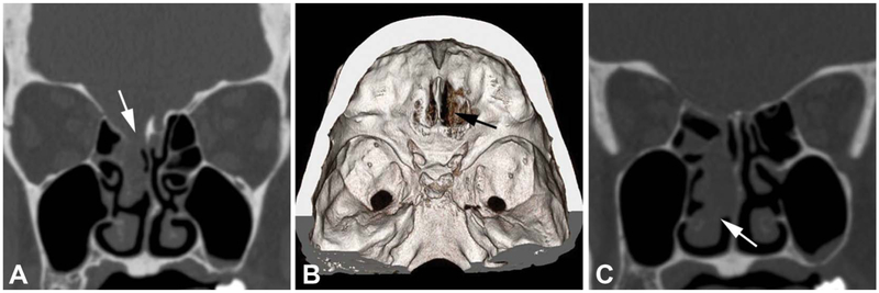
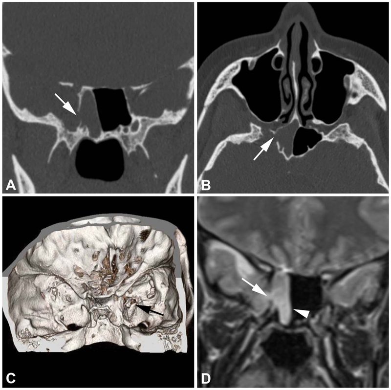
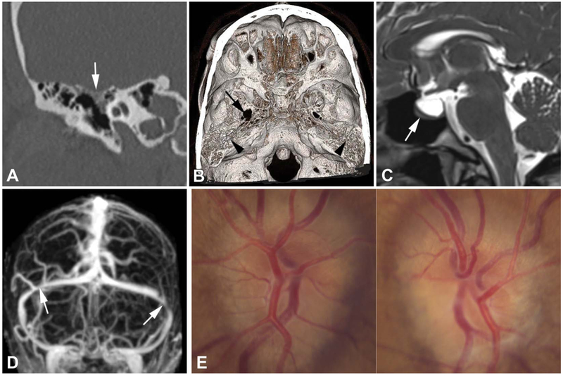
Subsequently, neuroimaging identifies the origin of the leak by localizing the osseous dehiscence (15). Noncontrast high-resolution computed tomography (CT) is the first-line imaging technique, with a sensitivity of about 90% and a specificity of 100% (17). Suggestive findings include one or multiple defect(s) of the skull base associated with an air–fluid level or opacification of adjacent cavities (paranasal sinuses, middle ear, and mastoid air cells) (17). The defect most often involves the ethmoid (Fig. 1A, B) and the sphenoid (Fig. 2A–C) bones in anterior CSF leaks, and the tegmen tympani and mastoidum of the temporal bone in lateral CSF leaks (Fig. 3A, B) (18,19). In patients with multiple defects or a negative CT, intrathecal administration of contrast with CT or magnetic resonance cisternography may help identify the site of the leak. Meningocele or meningoencephalocele also are frequently reported (Figs. 1C, 2D) (20–25). Brain MRI is useful in the differentiation of herniated intracranial contents from sequestered secretions within the contiguous cavities. In addition, brain MRI provides imaging findings consistent with long-standing intracranial hypertension (Fig. 3C, D) (19,26).
FIG. 1.
A 62-year-old woman with a BMI of 36 kg/m2 has a 4-year history of right-sided CSF rhinorrhea. A. Coronal computed tomography (CT) shows an osseous defect (arrow) of the right cribriform plate. B. Volume-rendered CT image, endocranial view, demonstrates the osseous defect (arrow). C. Coronal CT image (anterior to slice depicted in A) shows a meningoencephalocele (arrow) abutting the roof of the inferior turbinate. BMI, body mass index; CSF, cerebrospinal fluid.
FIG. 2.
A 46-year-old woman with a 20-year history of idiopathic intracranial hypertension and a BMI of 45 kg/m2 had placement of a ventriculoperitoneal shunt. Coronal (A) and axial (B) CT scans reveal an osseous defect (arrows) of the posterolateral wall of the right sphenoid sinus. C. Volume-rendered CT image, endocranial view, also shows the osseous defect (arrow). D. Coronal T2 MRI demonstrates a right temporal lobe meningoencephalocele (arrowhead) herniating through a bony dehiscence (arrow). BMI, body mass index; CT, computed tomography.
FIG. 3.
A 43-year-old woman has a history of fullness of her right ear, headaches, transient visual obscurations, and pulsatile tinnitus. Myringotomy yielded CSF otorrhea. BMI is 32 kg/m2. A. Coronal CT shows an osseous defect (arrow) of the right tegmen tympani and mastoidum. B. With volume-rendered CT image, endocranial view, the osseous defect cannot be localized. There is a moth-eaten appearance and thinning of the tegmen tympani bilaterally (arrowheads) and an eroded and widened right foramen ovale (arrow). C. Sagittal T2 MRI shows a partially empty sella (arrow). D. Posterior–inferior view of maximum intensity projection of postcontrast magnetic resonance venogram reveals bilateral transverse sinus stenosis (arrows). E. On funduscopy, there is bilateral papilledema. BMI, body mass index; CSF, cerebrospinal fluid; CT, computed tomography.
EVIDENCE FOR AN ASSOCIATION BETWEEN SPONTANEOUS CEREBROSPINAL FLUID LEAKS AND IDIOPATHIC INTRACRANIAL HYPERTENSION
Striking overlaps exist regarding demographic, clinical, and radiological characteristics between patients with IIH and those with spontaneous CSF leaks, suggesting that the 2 entities might indeed be related. In the following sections of this article, unless obtained from a review article, cited prevalence was obtained by pooling reported prevalence and rounding those results to the nearest multiple of 5.
Except for age, both groups share a similar demographic profile, with spontaneous CSF leaks mainly affecting obese patients (body mass index of approximately 36 kg/m2) and women (about 80%) (20,22,25,27–29). Interestingly, although spontaneous CSF leaks were initially considered rare, accounting for less than 10% of all causes of CSF leaks in early reports (9,10,30), recent case series have documented that CSF leaks are spontaneous in 30% of cases (22,31–34). This trend was confirmed by a survey that showed a doubling of the rate of surgical repair for spontaneous CSF leaks between 2002 and 2012 in the United States, paralleling the obesity epidemic, while the repair rate of nonspontaneous CSF leaks has remained unchanged (35). These reports corroborate our experience of increased numbers of patients referred with spontaneous CSF leak repair over the past 5 years. Patients with spontaneous CSF leaks are older (50 years) than typical IIH patients (36), perhaps because of delayed presentation. This is true since CSF leak may act as a “pressure release valve,” providing relief of signs and symptoms of IIH.
Many patients with spontaneous CSF leaks may harbor initial symptoms and signs suggestive of raised ICP. In the largest case series, headaches (60%) and pulsatile tinnitus (20%) are prominent, while diplopia occurred in 5% of cases (22,25,27,37,38). These rates are lower than those associated with classic IIH (90%, 60%, and 30%, respectively (39)), which is not surprising given the likelihood that the leak itself treats the intracranial hypertension and, therefore, relieves symptoms of raised ICP even in patients with IIH. This also would explain why papilledema has been uncommonly reported at presentation (2,4,14,40–42) and occasionally postoperatively (4,37,38). The few studies that have addressed papilledema before surgical repair have a pooled prevalence of approximately 5% (Fig. 3E) (22,27,36,42). However, it is unclear whether the prevalence of symptoms and signs of raised ICP in patients with spontaneous CSF leaks is lower than that of classic IIH or just underreported.
A few reports have suggested a temporal relationship between signs and symptoms of raised ICP and the onset of the leaks, as well as a direct effect of ICP lowering on the resolution of the leak. Development of spontaneous CSF leaks in patients with pre-existing, poorly controlled IIH has been reported (2–4,43–48). A few case reports and 1 study described a total of 22 patients with spontaneous CSF leaks without history of known IIH who developed symptoms or signs suggestive of raised ICP after surgical repair of the leak (4,7,49). Interestingly, a few case studies have included patients with spontaneous CSF leak whose leaks resolved after CSF shunt placement/revision (2,14,47) or after weight loss (46). However, because surgical repair of CSF leaks is the standard of treatment (50), reports regarding the nonconventional management of CSF leaks remain anecdotal.
Detection of radiographic signs of chronically raised ICP has never been addressed in patients with spontaneous CSF leaks. However, associated signs have often been reported as incidental findings, although with different prevalence than in classic IIH. Partial or complete empty sella, a radiographic sign of long-standing intracranial hypertension, has been described with a pooled prevalence in spontaneous CSF leak patients of 60% (13,21,22,25,27,28,33,34,37,38,51–54) vs 80% in patients with IIH (26). Radiographic signs related to mechanical deformation of the perioptic tissues secondary to raised ICP were found with a much lower prevalence in CSF leak patients as compared to those with IIH. Optic nerve tortuosity, dilation of the perioptic spaces, and flattened posterior sclera have been reported with a pooled prevalence of only 10% (13,28,38), 15% (13,28,38), and 5% (28,38), respectively, vs about 45%, 60%, and 65% in patients with classic IIH (26). Surprisingly, bilateral transverse venous stenosis, probably the most characteristic radiographic sign of long-standing raised ICP, has not been addressed in most studies, except for 2 reports (total of 3 patients) in which this finding was demonstrated (45,55).
An interesting phenomenon that lends further support to the association between raised ICP and spontaneous CSF leaks is the fact that leaks arising from the skull base are paradoxically often not associated with clinical and radiological signs of intracranial hypotension, unlike spinal CSF leaks (5). This might seem counterintuitive at first glance but may be explained by the fact that, in upright position, gravity induces a CSF pressure gradient with the CSF pressure being positive below the upper cervical spine and negative above this level (i.e., greater and lower than the atmospheric pressure, respectively). Consequently, a dehiscence at the level of the spine, resulting in CSF leak, will induce intracranial hypotension, whereas a dehiscence at the level of the skull base may not leak, as long as the CSF physiology remains normal (56). However, if the ICP rises above the atmospheric pressure at the level of the skull base where a dehiscence is present (such as in a patient with IIH), the patient will develop a CSF leak that will mitigate the ICP elevation, but no intracranial hypotension will ensue.
DIAGNOSING IDIOPATHIC INTRACRANIAL HYPERTENSION IN PATIENTS WITH SPONTANEOUS CEREBROSPINAL FLUID LEAKS
Clinicians face challenges when trying to determine whether a patient with spontaneous CSF leak may have IIH. Indeed, in most patients with IIH complicated by a leak, signs and symptoms of raised ICP may be obviated, and the diagnostic criteria for IIH (57) cannot be applied. In the few studies (22,24,25,28,58–62) suggesting the diagnosis of IIH in such patients, none provided details regarding the funduscopic examination. Although some authors did not specifically mention how the diagnosis was made (58,59), others solely relied on an ICP greater than 25 cm of water (28,60). A few studies used the 2002 (or earlier) Dandy criteria to make the diagnosis of IIH, but these criteria were often loosely applied, with missing CSF contents information (61) or ICP values (24). Some studies even applied nonstandard Dandy criteria for the diagnosis of IIH by substituting the strict criterion of elevated ICP with the presence of an empty sella when the ICP values ranged between 20 and 25 cm of water (22,25,62).
The 2002 Dandy criteria lack specificity, risking over-diagnosis of IIH, especially allowing for so-called IIH without papilledema (63). The more recent 2013 Dandy criteria (57) are much more constraining. Papilledema, 1 of the 5 major criteria, has seldom been reported in patients with spontaneous CSF leaks (27,36,38,42). In addition, in the small subset of IIH patients without papilledema, a CSF pressure greater than 25 cm of water is a mandatory criterion in addition to a sixth nerve palsy (57), whereas one would actually expect a lower ICP in patients with spontaneous CSF leaks because of the egress of CSF (36). Therefore, if neither papilledema nor ICP greater than 25 cm of water are present, a diagnosis of IIH cannot be made definitively in the majority of patients with spontaneous CSF leaks. Previous symptoms of intracranial hypertension in obese patients, findings of funduscopic changes suggestive of previous papilledema, or the presence of radiological signs of raised ICP are often the only way to suspect IIH as the cause of the leak.
CONTRIBUTION OF INTRACRANIAL PRESSURE TO THE PATHOPHYSIOLOGY OF SPONTANEOUS CEREBROSPINAL FLUID LEAKS
A better understanding of CSF dynamics in patients with spontaneous CSF leaks would provide substantial insight into the pathophysiology of this disorder. It has been suggested that persistent pulsatile high CSF pressure might exert eroding forces and remodeling at sites of intrinsic weakness, such as the thin bones of the skull base (61). The high rate of radiological signs of osseous erosion consistent with chronically elevated ICP, including point osseous erosion (arachnoid pits), meningoceles or encephaloceles, and empty sella, in patients with spontaneous CSF leaks supports this hypothesis (19). However, chronically elevated ICP is likely not the only factor, as only a minority of patients with chronically elevated ICP present with a spontaneous CSF leak, suggesting that these patients may have an anatomic predisposition such as abnormally thin or “weak” skull base bones. The extensive pneumatization of the sphenoid sinus with lateral expansion (so-called lateral recess of the sphenoid sinus) seen in 90% of patients with spontaneous CSF leaks originating from the sphenoid sinus as opposed to 20% of the general population supports this theory (64).
It also is possible that patients with spontaneous CSF leaks may have abnormal dura. The fact that meningoceles and meningoencephaloceles are found in 75% of patients with spontaneous CSF leak (13,21,22,24,25,34,37,52,53,58,64) compared with about 10% of classic IIH patients (65) supports this hypothesis.
Because high-pressure CSF flows along the path of least resistance, one could hypothesize the existence of 2 types of IIH patients: those with constitutionally fragile bones or other predisposition, more prone to CSF leaks; and those with normal bones and dura, more likely to present with classic IIH. The fact that, among patients with spontaneous CSF leaks, radiological signs of osseous erosion predominate over those related to mechanical deformation secondary to raised ICP supports this theory. Indeed, this discrepancy may mirror the apparent differential forces exerted by CSF pressure on the surrounding structures, more prominent on soft tissue for classic IIH compared with bone/dura for IIH-related CSF leaks.
Reliable measurements of ICP in patients with spontaneous CSF leaks would facilitate postoperative clinical decision-making after surgical repair. Despite numerous reports measuring preoperative and/or postoperative ICP in such patients, no consensus exists regarding ICP measurement in terms of timing, technique, and threshold above which patients are considered at high risk of postoperative intracranial hypertension. Indeed, not only was ICP not measured in numerous studies (8,21,32–34,52,64,66,67), but also those reporting such values have found inconsistent results. Although an average ICP greater than 25 cm of H2O either preoperatively and/or postoperatively was often demonstrated, individual results are highly variable, ranging from 2 to 60 cm of water (20,42), with some reports describing normal ICP (22,28,29,37,38,53,60). Several factors hinder the interpretation of these measurements. First, some studies combined preoperative and postoperative ICP results together (23,29,42), while the course of ICP should logically be altered after surgical repair, with ICP rising postoperatively (27,36,68,69). Second, although correct positioning of the patient and technique is paramount for reliable ICP measurements (70), many studies have provided few, if any, details regarding their methodology (2,4,27,41,46,47,54). Third, inconsistencies exist across studies with respect to the type of procedure (lumbar puncture (4,27,36,41,44,46,47,53,54) or lumbar drain (25,37,55,58)) and the timing of ICP measurement (before anesthesia (4,44,46,47) or after induction (25,36,53,58,68) for preoperative measurements, and between early (20,58,68) or late (28,38,61) postoperative period for postoperative measurements). Finally, CSF leaks might be patent or latent. Although the influence of whether CSF leaks are active at the time of the ICP measurement has never been addressed, higher ICP would be expected to produce continuous (patent) CSF leaks, whereas lower ICP would logically result in intermittent (latent) CSF leaks. Conversely, it is also possible that intermittent leaks may result from smaller holes and, therefore, may be associated with higher CSF pressure since less CSF escapes. The gold standard for measuring ICP is an intraventricular probe connected to an external transducer that measures the ICP continuously over a period (6). However, many authors provide a single ICP measurement by lumbar puncture or through a lumbar drain, which is only a snapshot of the CSF pressure. To the best of our knowledge, only one study of 12 patients (37) and one report of 2 cases (55) performed continuous measurements. All but one patient in these 2 studies had preoperative mean ICPs of less than 25 cm of water, with pooled data finding an ICP greater than 25 cm of water for only 10 ± 14% of the time preoperatively and 6 ± 12% postoperatively.
Another way to establish causality between IIH and spontaneous CSF leaks would require a prospective study assessing the prevalence of new-onset IIH after surgical repair of the CSF leak. However, a higher rate of success is achieved when ICP is actively controlled (see next section), and most clinicians routinely prescribe postoperative ICP-lowering agents such as acetazolamide or perform CSF shunting procedures in “high-risk” patients (e.g., history of IIH or CSF leak failure, high postoperative ICP) before or at the time of the leak repair (18), making a retrospective evaluation impossible. Recurrence of CSF leak after surgical repair is often considered a sign that ICP is abnormally elevated after the repair and, therefore, may indicate IIH as the cause of the leak. Ideally, systematic monitoring of ICP before and after surgical CSF leak repair during the early postoperative period should be performed. However, this rarely has been evaluated and the results are inconsistent, with some studies finding an increase in ICP of less than 10 cm of water postoperatively (including a total of 73 patients) (27,36,68), and others failing to demonstrate any ICP change (12 patients) (37) or even reporting a decrease in ICP postoperatively (16 patients) (58).
MANAGEMENT OF INTRACRANIAL PRESSURE AFTER CEREBROSPINAL FLUID LEAK REPAIR WHEN IDIOPATHIC INTRACRANIAL HYPERTENSION IS SUSPECTED
Unlike traumatic CSF leaks, spontaneous CSF leaks do not usually resolve spontaneously. Therefore, surgical repair is currently the standard of care to prevent complications (50) such as meningitis (71) or pneumocephalus (43). Surgical repair of anterior spontaneous CSF leaks usually is performed through endoscopic endonasal approaches, while those of lateral origin often require middle cranial fossa or transmastoid approaches.
Lumbar drain placement is an adjunct procedure occasionally used intraoperatively. Lumbar drainage of CSF presumably facilitates healing of the meninges during the first 2 or 3 postoperative days by releasing pressure at the surgical site and by preventing peaks of ICP (e.g., during Valsalva maneuvers) that would place the patient at risk of early recurrent leak (50). A lumbar drain also allows for intraoperative administration of fluorescein for better identification of the leaking site (50) and for ICP measurements on clamping of the drain to determine which patients may need long-term ICP management (42,53,54). However, its benefit remains controversial, especially given the risk of drainage-related complications including meningitis and pneumocephalus (72,73). Current recommendations are against routine use of lumbar drains due to inherent risks and a lack of evidence demonstrating a benefit (74).
Spontaneous CSF leaks may have a higher rate of failure after repair compared with those of traumatic origin (18). However, recent studies suggest that the rate for success of spontaneous CSF leak repair approximates that for traumatic CSF leaks, with an approximately 90% success rate when ICP is actively controlled postoperatively (18). In addition, closure of spontaneous CSF leaks has occasionally been reported only after an intervention aimed at decreasing the ICP (2,14,46,47). Although no consensus exists in the literature, a growing number of authors advocate for a more aggressive management of ICP postoperatively(18), such as with ICP-lowering agents (e.g., acetazolamide) or CSF diversion procedures. Endovascular venous stenting has been reported as adjunct intervention in 3 patients. This was performed because of failure to control ICP either with acetazolamide or CSF diversion surgery (45,55).
No study has prospectively examined the ideal postoperative ICP management, and only a few reports have provided their algorithm for long-term ICP management (22,42,54). Some authors have used acetazolamide during the postoperative period for at least 6 months, regardless of the ICP measurements (62), while others use it on a case-by-case basis (22,42,54); no details regarding the dose was reported in these studies, except one (1,000 mg daily (62)). A rationale does exist for prescribing acetazolamide, as 1 study (68) showed a decrease of ICP by 10 cm of water with its use after surgical repair of spontaneous CSF leaks. Similarly, the IIH Treatment Trial (75) showed a 10 cm of water decrease in ICP using 1–4 g/day of acetazolamide in IIH patients. However, acetazolamide’s impact on the recurrence of spontaneous CSF leaks has never been systematically studied. Similarly, a CSF shunting procedure (most often ventriculoperitoneal shunt) is sometimes performed at the time or after surgical repair in patients with refractory elevated ICP (24,29,36,54), but no study has systematically evaluated its benefits.
Surprisingly, although weight loss in obese patients is essential in IIH management, its impact on the prevention of spontaneous CSF leak recurrence remains unknown. Two anecdotal case reports (46,76) documented spontaneous closure of the CSF leak after bariatric surgery and subsequent weight loss. However, the need to achieve immediate ICP control to prevent early CSF leak recurrence in poorly controlled IIH patients limits the efficacy of weight loss over the short term (39). Despite the lack of evidence, weight loss is still recommended by some authors as an adjunct treatment after repair of a CSF leak (25,42,50).
CONCLUSIONS
IIH is becoming more widely recognized as a cause of spontaneous CSF leak. However, more than 20 years after the first case series of IIH-related CSF leaks was published (2), this causal relationship remains poorly characterized. Prospective studies have yet to be performed, and there is a lack of consensus regarding ICP management after surgical repair of the leak. Ultimately, revision of the IIH diagnostic criteria will likely include isolated spontaneous CSF leak with radiologic signs of raised ICP. In addition, systematic evaluation and follow-up by neuro-ophthalmologists of patients with spontaneous CSF leaks will help clarify the relationship between IIH and spontaneous CSF leaks.
Acknowledgments
Supported in part by an unrestricted departmental grant (Department of Ophthalmology) from Research to Prevent Blindness, Inc., New York, and by NIH/NEI core grant P30-EY06360 (Department of Ophthalmology). N. J. Newman received the Research to Prevent Blindness Lew R. Wasserman Merit Award. J. M. Levy is supported by the National Center for Advancing Translational Sciences of the National Institutes of Health under Award Numbers UL1TR002378 and KL2TR002381. The content is solely the responsibility of the authors and does not necessarily represent the official views of the National Institutes of Health.
Footnotes
The authors report no conflicts of interest.
REFERENCES
- 1.Ommaya AK, Di Chiro G, Baldwin M, Pennybacker JB. Non-traumatic cerebrospinal fluid rhinorrhoea. J Neurol Neurosurg Psychiatry. 1968;31:214–225. [DOI] [PMC free article] [PubMed] [Google Scholar]
- 2.Clark D, Bullock P, Hui T, Firth J. Benign intracranial hypertension: a cause of CSF rhinorrhoea. J Neurol Neurosurg Psychiatry. 1994;57:847–849. [DOI] [PMC free article] [PubMed] [Google Scholar]
- 3.Rosenfeld E, Dotan G, Kimchi TJ, Kesler A. Spontaneous cerebrospinal fluid otorrhea and rhinorrhea in idiopathic intracranial hypertension patients. J Neuroophthalmol. 2013;33:113–116. [DOI] [PubMed] [Google Scholar]
- 4.Pérez MA, Bialer OY, Bruce BB, Newman NJ, Biousse V. Primary spontaneous cerebrospinal fluid leaks and idiopathic intracranial hypertension. J Neuroophthalmol. 2013;33:330–337. [DOI] [PMC free article] [PubMed] [Google Scholar]
- 5.Schievink WI, Schwartz MS, Maya MM, Moser FG, Rozen TD. Lack of causal association between spontaneous intracranial hypotension and cranial cerebrospinal fluid leaks. J Neurosurg. 2012;116:749–754. [DOI] [PubMed] [Google Scholar]
- 6.Ducros A, Biousse V. Headache arising from idiopathic changes in CSF pressure. Lancet Neurol. 2015;14:655–668. [DOI] [PubMed] [Google Scholar]
- 7.Mokri B Intracranial hypertension after treatment of spontaneous cerebrospinal fluid leaks. Mayo Clin Proc. 2002;77:1241–1246. [DOI] [PubMed] [Google Scholar]
- 8.Badia L, Loughran S, Lund V. Primary spontaneous cerebrospinal fluid rhinorrhea and obesity. Am J Rhinol. 2001;15:117–119. [DOI] [PubMed] [Google Scholar]
- 9.Eljamel MS, Foy PM. Non-traumatic CSF fistulae: clinical history and management. Br J Neurosurg. 1991;5:275–279. [DOI] [PubMed] [Google Scholar]
- 10.Hubbard JL, McDonald TJ, Pearson BW, Laws ER. Spontaneous cerebrospinal fluid rhinorrhea: evolving concepts in diagnosis and surgical management based on the Mayo Clinic experience from 1970 through 1981. Neurosurgery. 1985;16:314–321. [DOI] [PubMed] [Google Scholar]
- 11.Shugar JM, Som PM, Eisman W, Biller HF. Non-traumatic cerebrospinal fluid rhinorrhea. Laryngoscope. 1981;91:114–120. [PubMed] [Google Scholar]
- 12.Brisman R, Hughes JE, Mount LA. Cerebrospinal fluid rhinorrhea. Arch Neurol. 1970;22:245–252. [DOI] [PubMed] [Google Scholar]
- 13.Silver RI, Moonis G, Schlosser RJ, Bolger WE, Loevner LA. Radiographic signs of elevated intracranial pressure in idiopathic cerebrospinal fluid leaks: a possible presentation of idiopathic intracranial hypertension. Am J Rhinol. 2007;21:257–261. [DOI] [PubMed] [Google Scholar]
- 14.Darouassi Y, Mliha Touati M, Chihani M, Akhaddar A, Ammar H, Bouaity B. Spontaneous cerebrospinal fluid leak of the sphenoid sinus mimicking allergic rhinitis, and managed successfully by a ventriculoperitoneal shunt: a case report. J Med Case Rep. 2016;10:308. [DOI] [PMC free article] [PubMed] [Google Scholar]
- 15.Wang EW, Vandergrift WA, Schlosser RJ. Spontaneous CSF leaks. Otolaryngol Clin North Am. 2011;44:845–vii. [DOI] [PubMed] [Google Scholar]
- 16.Oakley GM, Alt JA, Schlosser RJ, Harvey RJ, Orlandi RR. Diagnosis of cerebrospinal fluid rhinorrhea: an evidence-based review with recommendations. Int Forum Allergy Rhinol. 2016;6:8–16. [DOI] [PubMed] [Google Scholar]
- 17.Lloyd KM, DelGaudio JM, Hudgins PA. Imaging of skull base cerebrospinal fluid leaks in adults. Radiology. 2008;248:725–736. [DOI] [PubMed] [Google Scholar]
- 18.Teachey W, Grayson J, Cho DY, Riley KO, Woodworth BA. Intervention for elevated intracranial pressure improves success rate after repair of spontaneous cerebrospinal fluid leaks. Laryngoscope. 2017;127:2011–2016. [DOI] [PubMed] [Google Scholar]
- 19.Alonso RC, de la Peña MJ, Caicoya AG, Rodriguez MR, Moreno EA, de Vega Fernandez VM. Spontaneous skull base meningoencephaloceles and cerebrospinal fluid fistulas. Radiographics. 2013;33:553–570. [DOI] [PubMed] [Google Scholar]
- 20.Woodworth BA, Prince A, Chiu AG, Cohen NA, Schlosser RJ, Bolger WE, Kennedy DW, Palmer JN. Spontaneous CSF leaks: a paradigm for definitive repair and management of intracranial hypertension. Otolaryngol Head Neck Surg. 2008;138:715–720. [DOI] [PubMed] [Google Scholar]
- 21.Goddard JC, Meyer T, Nguyen S, Lambert PR. New considerations in the cause of spontaneous cerebrospinal fluid otorrhea. Otol Neurotol. 2010;31:940–945. [DOI] [PubMed] [Google Scholar]
- 22.Seth R, Rajasekaran K III, Luong A, Benninger MS, Batra PS. Spontaneous CSF leaks: factors predictive of additional interventions. Laryngoscope. 2010;120:2141–2146. [DOI] [PubMed] [Google Scholar]
- 23.Illing E, Schlosser RJ, Palmer JN, Curé J, Fox N, Woodworth BA. Spontaneous sphenoid lateral recess cerebrospinal fluid leaks arise from intracranial hypertension, not Sternberg’s canal. Int Forum Allergy Rhinol. 2014;4:246–250. [DOI] [PubMed] [Google Scholar]
- 24.Campbell RG, Farquhar D, Zhao N, Chiu AG, Adappa ND, Palmer JN. Cerebrospinal fluid rhinorrhea secondary to idiopathic intracranial hypertension: long-term outcomes of endoscopic repairs. Am J Rhinol Allergy. 2016;30:294–300. [DOI] [PubMed] [Google Scholar]
- 25.Martínez-Capoccioni G, Serramito-García R, Martín-Bailón M, García-Allut A, Martín-Martín C. Spontaneous cerebrospinal fluid leaks in the anterior skull base secondary to idiopathic intracranial hypertension. Eur Arch Otorhinolaryngol. 2017;274:2175–2181. [DOI] [PubMed] [Google Scholar]
- 26.Bidot S, Saindane AM, Peragallo JH, Bruce BB, Newman NJ, Biousse V. Brain imaging in idiopathic intracranial hypertension. J Neuroophthalmol. 2015;35:400–411. [DOI] [PubMed] [Google Scholar]
- 27.Yang Z, Wang B, Wang C, Liu P. Primary spontaneous cerebrospinal fluid rhinorrhea: a symptom of idiopathic intracranial hypertension? J Neurosurg. 2011;115:165–170. [DOI] [PubMed] [Google Scholar]
- 28.Allen KP, Perez CL, Kutz JW, Gerecci D, Roland PS, IsaacsonB. Elevated intracranial pressure in patients with spontaneous cerebrospinal fluid otorrhea. Laryngoscope. 2014;124:251–254. [DOI] [PubMed] [Google Scholar]
- 29.Vivas EX, Mccall A, Raz Y, Fernandez-Miranda JC, Gardner P, Hirsch BE. ICP, BMI, surgical repair, and CSF diversion in patients presenting with spontaneous CSF otorrhea. Otol Neurotol. 2014;35:344–347. [DOI] [PubMed] [Google Scholar]
- 30.Spetzler RF, Wilson CB. Management of recurrent CSF rhinorrhea of the middle and posterior fossa. J Neurosurg. 1978;49:393–397. [DOI] [PubMed] [Google Scholar]
- 31.Lopatin AS, Kapitanov DN, Potapov AA. Endonasal endoscopic repair of spontaneous cerebrospinal fluid leaks. Arch Otolaryngol Head Neck Surg. 2003;129:859–863. [DOI] [PubMed] [Google Scholar]
- 32.Carrau RL, Snyderman CH, Kassam AB. The management of cerebrospinal fluid leaks in patients at risk for high-pressure hydrocephalus. Laryngoscope. 2005;115:205–212. [DOI] [PubMed] [Google Scholar]
- 33.Mirza S, Thaper A, McClelland L, Jones NS. Sinonasal cerebrospinal fluid leaks: management of 97 patients over 10 years. Laryngoscope. 2005;115:1774–1777. [DOI] [PubMed] [Google Scholar]
- 34.Quatre R, Attye A, Righini C, Reyt E, Giai J, Schmerber S, Karkas A. Spontaneous cerebrospinal fluid rhinorrhea: association with body weight and imaging data. J Neurol Surg B Skull Base. 2017;78:419–424. [DOI] [PMC free article] [PubMed] [Google Scholar]
- 35.Nelson RF, Gantz BJ, Hansen MR. The rising incidence of spontaneous cerebrospinal fluid leaks in the United States and the association with obesity and obstructive sleep apnea. Otol Neurotol. 2015;36:476–480. [DOI] [PubMed] [Google Scholar]
- 36.Aaron G, Doyle J, Vaphiades MS, Riley KO, Woodworth BA. Increased intracranial pressure in spontaneous CSF leak patients is not associated with papilledema. Otolaryngol Head Neck Surg. 2014;151:1061–1066. [DOI] [PubMed] [Google Scholar]
- 37.Reh DD, Gallia GL, Ramanathan M, Solomon D, Moghekar A, Ishii M, Lane AP. Perioperative continuous cerebrospinal fluid pressure monitoring in patients with spontaneous cerebrospinal fluid leaks: presentation of a novel technique. Am J Rhinol Allergy. 2010;24:238–243. [DOI] [PubMed] [Google Scholar]
- 38.Jiang ZY, McLean C, Perez C, Barnett S, Friedman D, Tajudeen BA, Batra PS. Surgical outcomes and postoperative management in spontaneous cerebrospinal fluid rhinorrhea. J Neurol Surg B Skull Base. 2018;79:193–199. [DOI] [PMC free article] [PubMed] [Google Scholar]
- 39.Bidot S, Bruce BB. Update on the diagnosis and treatment of idiopathic intracranial hypertension. Semin Neurol. 2015;35:527–538. [DOI] [PubMed] [Google Scholar]
- 40.Ozveren MF, Kaplan M, Topsakal C, Bilge T, Erol FS, Celiker H, Akdemir I, Uchida K. Spontaneous cerebrospinal fluid rhinorrhea associated with chronic renal failure—case report. Neurol Med Chir (Tokyo). 2001;41:313–317. [DOI] [PubMed] [Google Scholar]
- 41.Saifudheen K, Gafoor A, Arun G, Abdurahiman P, Jose J. Idiopathic intracranial hypertension presenting as CSF rhinorrhea. Ann Indian Acad Neurol. 2010;13:72–73. [DOI] [PMC free article] [PubMed] [Google Scholar]
- 42.Fyrmpas G, Konstantinidis I, Selviaridis P, Constantinidis J. Management of spontaneous cerebrospinal fluid leaks of the sphenoid sinus: our experience. J Laryngol Otol. 2014;128:797–802. [DOI] [PubMed] [Google Scholar]
- 43.Baba M, Tarar O, Syed A. A rare case of spontaneous pneumocephalus associated with nontraumatic cerebrospinal fluid leak. Case Rep Neurol Med. 2016;2016:1828461. [DOI] [PMC free article] [PubMed] [Google Scholar]
- 44.Camras LR, Ecanow JS, Abood CA. Spontaneous cerebrospinal fluid rhinorrhea in a patient with pseudotumor cerebri. J Neuroimaging. 1998;8:41–42. [DOI] [PubMed] [Google Scholar]
- 45.Owler BK, Allan R, Parker G, Besser M. Pseudotumour cerebri, CSF rhinorrhoea and the role of venous sinus stenting in treatment. Br J Neurosurg. 2003;17:79–83. [DOI] [PubMed] [Google Scholar]
- 46.Rudnick E, Sismanis A. Pulsatile tinnitus and spontaneous cerebrospinal fluid rhinorrhea: indicators of benign intracranial hypertension syndrome. Otol Neurotol. 2005;26:166–168. [DOI] [PubMed] [Google Scholar]
- 47.Ransom ER, Komotar RJ, Mocco J, Connolly ES, Mullins KJ. Shunt failure in idiopathic intracranial hypertension presenting with spontaneous cerebrospinal fluid leak. J Clin Neurosci. 2006;13:598–602. [DOI] [PubMed] [Google Scholar]
- 48.Suryadevara AC, Fattal M, Woods CI. Nontraumatic cerebrospinal fluid rhinorrhea as a result of pseudotumor cerebri. Am J Otolaryngol. 2007;28:242–246. [DOI] [PubMed] [Google Scholar]
- 49.Schlosser RJ, Wilensky EM, Grady MS, Bolger WE. Elevated intracranial pressures in spontaneous cerebrospinal fluid leaks. Am J Rhinol. 2003;17:191–195. [PubMed] [Google Scholar]
- 50.Lobo BC, Baumanis MM, Nelson RF. Surgical repair of spontaneous cerebrospinal fluid (CSF) leaks: a systematic review. Laryngoscope Investig Otolaryngol. 2017;2:215–224. [DOI] [PMC free article] [PubMed] [Google Scholar]
- 51.Schlosser RJ, Bolger WE. Spontaneous nasal cerebrospinal fluid leaks and empty sella syndrome: a clinical association. Am J Rhinol. 2003;17:91–96. [PubMed] [Google Scholar]
- 52.Prichard CN, Isaacson B, Oghalai JS, Coker NJ, Vrabec JT. Adult spontaneous CSF otorrhea: correlation with radiographic empty sella. Otolaryngol Head Neck Surg. 2006;134:767–771. [DOI] [PubMed] [Google Scholar]
- 53.Kenning TJ, Willcox TO, Artz GJ, Schiffmacher P, Farrell CJ, Evans JJ. Surgical management of temporal meningoencephaloceles, cerebrospinal fluid leaks, and intracranial hypertension: treatment paradigm and outcomes. Neurosurg Focus. 2012;32:E6. [DOI] [PubMed] [Google Scholar]
- 54.Chaaban MR, Illing E, Riley KO, Woodworth BA. Spontaneous cerebrospinal fluid leak repair: a five-year prospective evaluation. Laryngoscope. 2014;124:70–75. [DOI] [PubMed] [Google Scholar]
- 55.Iyer RR, Solomon D, Moghekar A, Goodwin CR, Stewart CM, Ishii M, Gailloud P, Gallia GL. Venous sinus stenting in the management of patients with intracranial hypertension manifesting with skull base cerebrospinal fluid leaks. World Neurosurg. 2017;106:103–112. [DOI] [PubMed] [Google Scholar]
- 56.Kranz PG, Gray L, Amrhein TJ. Spontaneous intracranial hypotension: 10 myths and misperceptions. Headache. 2018;58:948–959. [DOI] [PubMed] [Google Scholar]
- 57.Friedman DI, Liu GT, Digre KB. Revised diagnostic criteria for the pseudotumor cerebri syndrome in adults and children. Neurology. 2013;81:1159–1165. [DOI] [PubMed] [Google Scholar]
- 58.Ramakrishnan VR, Suh JD, Chiu AG, Palmer JN. Reliability of preoperative assessment of cerebrospinal fluid pressure in the management of spontaneous cerebrospinal fluid leaks and encephaloceles. Int Forum Allergy Rhinol. 2011;1:201–205. [DOI] [PubMed] [Google Scholar]
- 59.Markou K, Goudakos J, Franco-Vidal V, Vergnolles V, Vignes JR, Darrouzet V. Spontaneous osteodural defects of the temporal bone: diagnosis and management of 12 cases. Am J Otolaryngol. 2011;32:135–140. [DOI] [PubMed] [Google Scholar]
- 60.Brainard L, Chen DA, Aziz KM, Hillman TA. Association of benign intracranial hypertension and spontaneous encephalocele with cerebrospinal fluid leak. Otol Neurotol. 2012;33:1621–1624. [DOI] [PubMed] [Google Scholar]
- 61.Schlosser RJ, Woodworth BA, Wilensky EM, Grady MS, Bolger WE. Spontaneous cerebrospinal fluid leaks: a variant of benign intracranial hypertension. Ann Otol Rhinol Laryngol. 2006;115:495–500. [DOI] [PubMed] [Google Scholar]
- 62.Martínez-Capoccioni G, Serramito-García R, Huertas-Pardo B, García-Allut A, Martín-Martín C. Spontaneous cerebrospinal fluid leaks in the anterior skull base: a surgical challenge. J Laryngol Otol. 2015;129:358–364. [DOI] [PubMed] [Google Scholar]
- 63.Fisayo A, Bruce BB, Newman NJ, Biousse V. Overdiagnosis of idiopathic intracranial hypertension. Neurology. 2016;86:341–350. [DOI] [PMC free article] [PubMed] [Google Scholar]
- 64.Shetty PG, Shroff MM, Fatterpekar GM, Sahani DV, Kirtane MV. A retrospective analysis of spontaneous sphenoid sinus fistula: MR and CT findings. AJNR Am J Neuroradiol. 2000;21:337–342. [PMC free article] [PubMed] [Google Scholar]
- 65.Bialer OY, Rueda MP, Bruce BB, Newman NJ, Biousse V, Saindane AM. Meningoceles in idiopathic intracranial hypertension. AJR Am J Roentgenol. 2014;202:608–613. [DOI] [PMC free article] [PubMed] [Google Scholar]
- 66.Scurry WC, Ort SA, Peterson WM, Sheehan JM, Isaacson JE. Idiopathic temporal bone encephaloceles in the obese patient. Otolaryngol Head Neck Surg. 2007;136:961–965. [DOI] [PubMed] [Google Scholar]
- 67.Dunn CJ, Alaani A, Johnson AP. Study on spontaneous cerebrospinal fluid rhinorrhoea: its aetiology and management. J Laryngol Otol. 2005;119:12–15. [DOI] [PubMed] [Google Scholar]
- 68.Chaaban MR, Illing E, Riley KO, Woodworth BA. Acetazolamide for high intracranial pressure cerebrospinal fluid leaks. Int Forum Allergy Rhinol. 2013;3:718–721. [DOI] [PubMed] [Google Scholar]
- 69.Aaron GP, Illing E, Lambertsen Z, Ritter M, Middlebrooks EH, Cure J, Cho DY, Riley KO, Woodworth BA. Enlargement of Meckel’s cave in patients with spontaneous cerebrospinal fluid leaks. Int Forum Allergy Rhinol. 2017;7:421–424. [DOI] [PubMed] [Google Scholar]
- 70.Lee SC, Lueck CJ. Cerebrospinal fluid pressure in adults. J Neuroophthalmol. 2014;34:278–283. [DOI] [PubMed] [Google Scholar]
- 71.Daudia A, Biswas D, Jones NS. Risk of meningitis with cerebrospinal fluid rhinorrhea. Ann Otol Rhinol Laryngol. 2007;116:902–905. [DOI] [PubMed] [Google Scholar]
- 72.Nelson RF, Roche JP, Gantz BJ, Hansen MR. Middle cranial fossa (MCF) approach without the use of lumbar drain for the management of spontaneous cerebral spinal fluid (CSF) leaks. Otol Neurotol. 2016;37:1625–1629. [DOI] [PubMed] [Google Scholar]
- 73.Caballero N, Bhalla V, Stankiewicz JA, Welch KC. Effect of lumbar drain placement on recurrence of cerebrospinal rhinorrhea after endoscopic repair. Int Forum Allergy Rhinol. 2012;2:222–226. [DOI] [PubMed] [Google Scholar]
- 74.Oakley GM, Orlandi RR, Woodworth BA, Batra PS, Alt JA. Management of cerebrospinal fluid rhinorrhea: an evidence-based review with recommendations. Int Forum Allergy Rhinol. 2016;6:17–24. [DOI] [PubMed] [Google Scholar]
- 75.NORDIC Idiopathic Intracranial Hypertension Study Group Writing Committee, Wall M, McDermott MP, Kieburtz KD, Corbett JJ, Feldon SE, Friedman DI, Katz DM, Keltner JL, Schron EB, Kupersmith MJ. Effect of acetazolamide on visual function in patients with idiopathic intracranial hypertension and mild visual loss: the idiopathic intracranial hypertension treatment trial. JAMA. 2014;311:1641–1651. [DOI] [PMC free article] [PubMed] [Google Scholar]
- 76.Stangherlin P, Ledeghen S, Scordidis V, Rubay R. Benign intracranial hypertension with recurrent spontaneous cerebrospinal fluid rhinorrhoea treated by laparoscopic gastric banding. Acta Chir Belg. 2008;108:616–618. [DOI] [PubMed] [Google Scholar]


