FIG. 3.
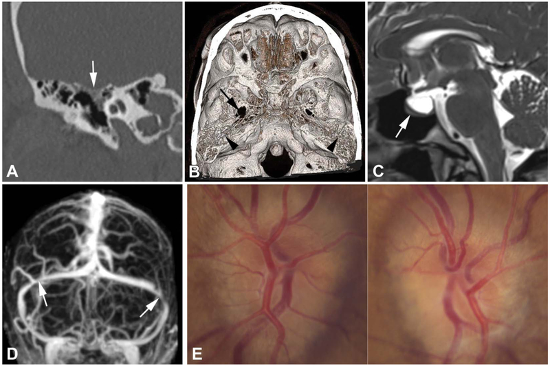
A 43-year-old woman has a history of fullness of her right ear, headaches, transient visual obscurations, and pulsatile tinnitus. Myringotomy yielded CSF otorrhea. BMI is 32 kg/m2. A. Coronal CT shows an osseous defect (arrow) of the right tegmen tympani and mastoidum. B. With volume-rendered CT image, endocranial view, the osseous defect cannot be localized. There is a moth-eaten appearance and thinning of the tegmen tympani bilaterally (arrowheads) and an eroded and widened right foramen ovale (arrow). C. Sagittal T2 MRI shows a partially empty sella (arrow). D. Posterior–inferior view of maximum intensity projection of postcontrast magnetic resonance venogram reveals bilateral transverse sinus stenosis (arrows). E. On funduscopy, there is bilateral papilledema. BMI, body mass index; CSF, cerebrospinal fluid; CT, computed tomography.

