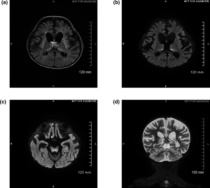
Figure 1.

Cerebral magnetic resonance imaging performed at 4 years of age. Axial (a–c) and coronal planes (d). (a) FLAIR sequence shows hypersignals of the basal ganglia and thalami. (b) and (c) reveal hypersignals in thalami and brainstem on diffusion‐weighted imaging. (d) T2‐weighted imaging indicates global cortico‐subcortical atrophy. FLAIR, fluid attenuated recovery
