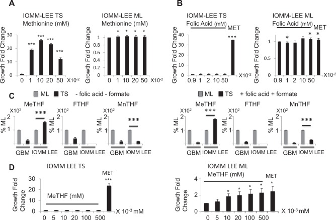
Fig. 6. Contrary to the glioblastoma cell lines, folic acid, and 5-methyltetrahydrofolate fail to promote tumor spheres formation in the meningioma cell line IOMM-LEE.
a IOMM-LEE TS are methionine dependent. Methionine, up to 0.1 mM increases TS growth. Cells are cultured in methionine free media supplemented with the indicated amounts of exogenous methionine. Samples are compared to cells grown with no exogenous methionine addition, n = 9. b Folic Acid does not rescue IOMM-LEE TS formation in methionine depleted media. Cells are grown in methionine free DMEM which already contains 0.009 mM folic acid, then more folic acid is added to the media to reach the indicated final concentrations. The MET sample is used as a reference and represents cells grown in DMEM containing 0.01 mM methionine. Samples are compared to cells grown in 0.009 mM folic acid, n = 9. c 5-Methyltetrahydrofolate level is higher in IOMM-LEE TS than ML. 10-Formyltetrahydrofolate and 5,10-methenyltetrahydrofolate exhibit lower concentrations in IOMM-LEE TS compared to IOMM-LEE ML cells, however, 5-methyltetrahydrofolate is more elevated in TS than in ML cells. Addition of folic acid and formate keeps these results unchanged. Folate isoforms are averaged in all 4 glioblastoma cell lines and the mean is represented. « + » sign indicates that folic acid and formate were added to the media and «−» sign that they were not added, n = 3. d 5-Methyltetrahydrofolate does not restore U251 TS formation, n = 3. Cells are grown in methionine free media then 5-methyltetrahydrofolate is added to reach the indicated concentrations. The MET sample is used as a reference and represents cells grown in DMEM containing 0.01 mM methionine. *p < 0.05, **p < 0.01, ***p < 0.001, MeTHF = 5-methyltetrahydrofolate, FTHF = 10-formyltetrahydrofolate, MnTHF = 5,10-methenyl tetrahydrofolate, ML = monolayer cells, TS = tumor spheres

