Fig. 7.
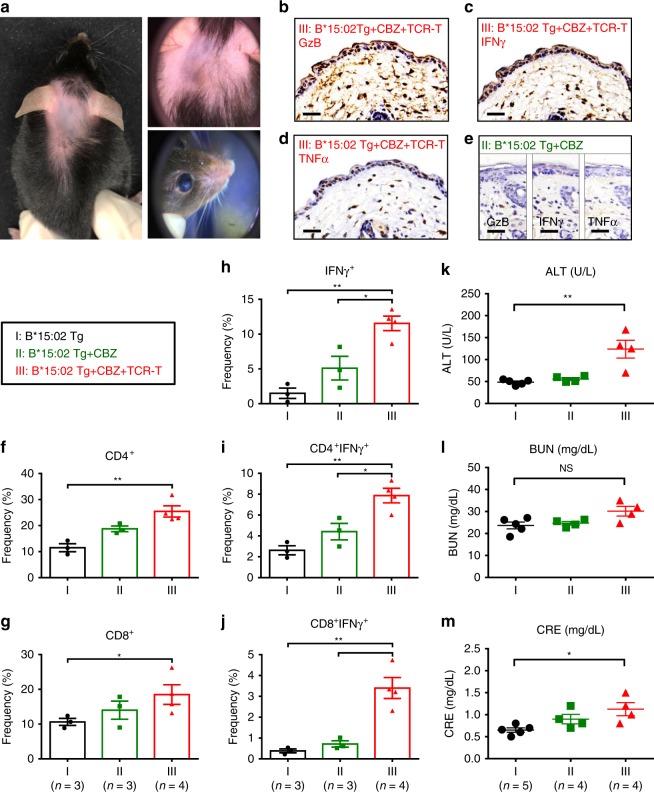
Adoptive transfer of TCR-T lymphocytes to HLA-B*15:02 transgenic mice. HLA-B*15:02 transgenic mice were assigned to the group (I) the vehicle controls (n = 5); group (II) given carbamazepine daily (328 mg/kg/day) by oral gavage (n = 4); and group (III) given carbamazepine daily and adoptive transfer of the public TCR-T (n = 4). Photographs, biopsies of the affected skin, and peripheral blood were obtained. a Representative photos of the affected skin and eyes of the group III mice are shown. b–e Immunohistochemistry staining of the skin biopsies of the mice. Compared with the group II, the group III mice showed augmented expression of cytotoxic protein granzyme B (GzB), and inflammatory cytokines, including IFNγ and TNFα. The results are representative of three independent experiments. Scale bar indicates 100 µm. f–j The frequencies of CD4+, CD8+, and/or IFN+ lymphocytes in the peripheral blood of the mice group I (n = 3), group II (n = 3), and group III (n = 4). The results are expressed as mean ± s.e.m. with each dot representing the data of an individual mouse. Statistical analysis was performed using an unpaired, two-tailed Student’s t test. k–m The plasma levels of ALT (alanine aminotransferase), BUN (blood urea nitrogen), and CRE (creatinine) were determined in the mice group I (n = 5), group II (n = 4), and group III (n = 4). The levels of each parameter are plotted as the mean ± s.e.m. with each dot representing the data of an individual mouse. Statistical analysis was performed using an unpaired, two-tailed Student’s t test. *P < 0.05; **P < 0.01

