Figure 3.
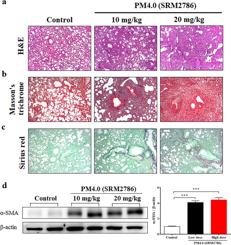
PM4.0 exposure increases the activation of pulmonary inflammation and fibrosis in NF-κB-luciferase+/+ transgenic mice. (a) Morphologic features of mouse lung inflammation indicated by hematoxylin and eosin (H&E) staining. The thicknesses of the bronchial wall, epithelial layer and smooth muscle in the airways of the PM4.0 groups were higher than those of the control group. Representative photomicrographs showing H&E staining (magnification, 100x). (b) Collagen deposition in the lung tissue of mice was observed by Masson’s trichrome staining. Hypertrophy, dense collagen bundles, and increased collagen deposition were present in the PM4.0 groups compared with the control group. Representative photomicrographs showing Masson’s trichrome staining (magnification, 100x). (c) Collagen fibers in the lung tissue of mice were observed by Sirius red staining. More collagen fibers were present in the PM4.0 groups than in the with control group. Representative photomicrographs showing Sirius red staining (magnification, 100x). The scale bars in all images are 100 μm. (d) Changes in the protein expression level of α-SMA in different groups normalized to the internal control, β-actin. PM4.0 exposure increased the α-SMA expression level in the PM4.0 groups compared with that in the control group. Representative images showing the protein expression levels assayed by Western blotting. n = 8 per group. Data are expressed as the mean ± SD. ***p < 0.001 compared to the control group.

