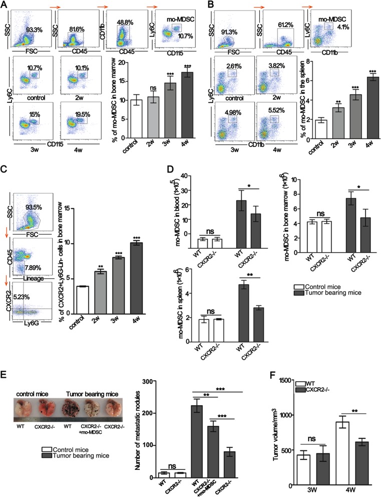
Fig. 1. A lack of CXCR2 leads to reduced mo-MDSCs accumulation and delayed tumor progression.
a, b The percentage of mo-MDSCs in the bone marrow and spleen of tumor-bearing mice was analyzed by flow cytometry. The samples were analyzed on the second, third, and week 4th of tumor-bearing. c The CXCR2 positive cells in bone marrow were analyzed by flow cytometry. The mice were treated as described in a, b. d Absolute number of mo-MDSCs in the blood, bone marrow, and spleen of control mice or mice bearing tumors after 3 weeks. e The number of metastatic foci was detected in the lungs of WT control mice, CXCR2−/− control mice, 2-week WT, 2-week CXCR2−/−, and adoptively transferred mo-MDSCs in two-week CXCR2−/− tumor-bearing mice. All mice were sacrificed at 2 weeks after a tail vein injection of B16F10 cells. The transferred mo-MDSCs were isolated from the blood of WT tumor-bearing mice. f B16F10 cells were subcutaneously inoculated into WT or CXCR2−/− mice, and the size of tumors was measured at the indicated time points. Bars represent the mean ± SD of five independent experiments. A one-way ANOVA with repeated measures followed by a Dunnett’s post hoc test or a two-way ANOVA followed by Holm-Sidak’s post hoc test were used to determine the level of statistical significance (*p < 0.05; **p < 0.01; and ***p < 0.001; ns, not significant)

