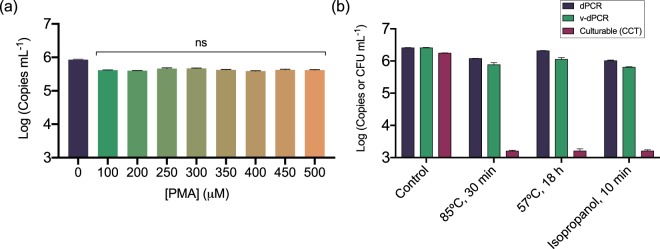
Figure 3.
Effect of PMA concentration and the E. amylovora killing method on the exclusion of false-positive signals by v-dPCR. Apple branch macerates prepared in AMB were inoculated with 106 E. amylovora heat-killed cells mL−1 (85 °C for 30 min), and treated with PMA concentrations ranging from 0–500 μM before DNA extraction and dPCR (a). E. amylovora live cells (103 CFU mL−1) were mixed with dead cells (106 cell mL−1) obtained either by heating at 85 °C for 30 min, heating at 57 °C for 18 h, or exposure to 70% isopropanol for 10 min. Samples were then analyzed by dPCR, v-dPCR (treatment with 100 μM PMA) and plate counts (b). D-PCRs were performed using the same primers and probe as Pirc et al.31. Represented data are mean values of experiments performed in triplicate. Error bars show the SD.

