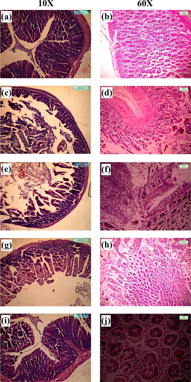
Figure 11.

H&E staining of mice colon. (a,b) Normal colon at 10× and 60× magnifications; (c,d) Initiation of colon adenocarcinoma after treatment with DMH for a 7 weeks (at 10× and 60× magnifications respectively); (e,f) Development of colon adenocarcinoma after completion of treatment regimen with DMH (12 weeks) (at 10× and 60× magnification respectively); (g,h) Improvement of cancerous region after completion of treatment of colorectal cancer mice treated with 2c-NP (9 weeks) (at 10× and 60× magnifications respectively). (I,j) Normal colon after treatment with 2c-NP (at 10× and 60× magnifications respectively). Scale bar used in 10× and 60× magnification panels represent 200 µm and 20 µm respectively.
