Figure 1.
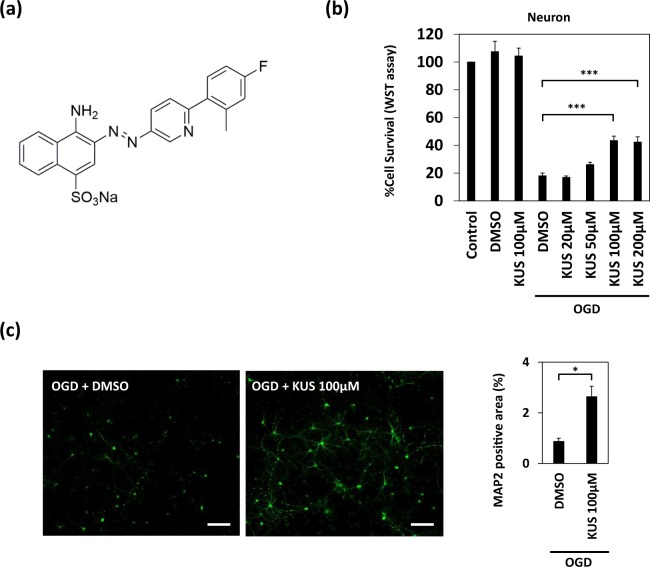
KUS121 protects primary cortical neurons under oxygen glucose deprivation (OGD). (a) The chemical structure of KUS121, an ATPase inhibitor of valosin-containing protein (VCP). (b) Cell viability measured by WST (water-soluble tetrazolium salt) assay. Primary cortical neurons from the rats were exposed to vehicle (dimethyl sulfoxide, DMSO) or KUS121 under control conditions or under OGD for 2 h (n = 4; ***p < 0.001). (c) Left: MAP2 stained rat primary cortical neurons after OGD. Primary neurons were exposed to OGD for 1.5 h in the presence of vehicle (DMSO) or 100 μM KUS121. Scale bar: 100 μm. Right: Graph showing the ratio of MAP2-positive area to the total image area (n = 4; *p < 0.05).

