Figure 4.

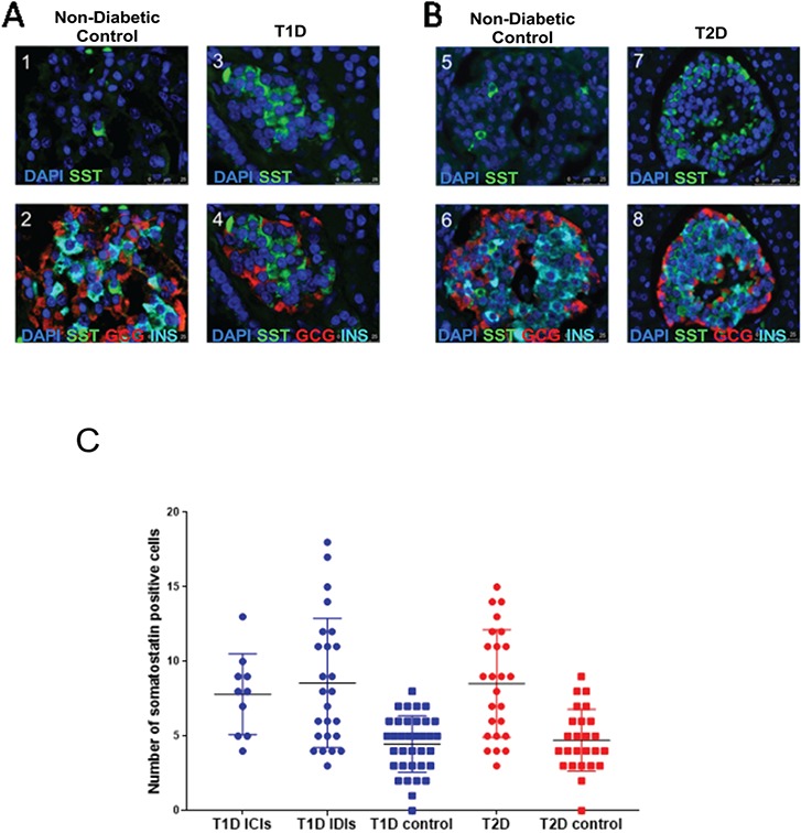
Hormone staining patterns of donor islets from controls or individuals with T2D. (A) Panels 1–4 are representative immunofluorescence images from human donor pancreatic tissue from controls (panels 1 and 2) and from cases of T1D compared to matched controls (panels 3 and 4). The identity of the antibody used to stain is indicated in each panel. (B) Panels 5–8 are representative immunofluorescence images from human donor pancreatic tissue from controls (panels 5 and 6) and from cases of T2D compared to matched controls (panels 7 and 8). (C) Graph showing interquartile range from delta cell counts for individuals without diabetes and patients with either T1D or T2D. We found a significant increase in the number of SST-positive cells in patients with either T1D INS-containing islets and INS-deficient islets (P = 3.0 × 10⁻⁶) or T2D (P = 2.20× 10⁻⁴) compared to their respective controls.
