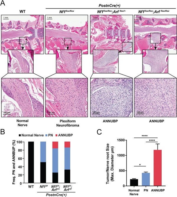
Figure 6.

Frequency and size of PN and ANNUBP arising from the cervicothoracic spine of Nf1/Arf mutant mice. (A) Coronal whole spinal sections revealing the spinal cord, DRG with associated proximal spinal nerve roots demonstrating PN and ANNUBP arising from the proximal nerve roots of Nf1flox/flox;PostnCre(+), Nf1flox/flox;Arfflox/+;PostnCre(+) and Nf1flox/flox;Arfflox/flox;PostnCre(+) mice. WT mice exhibited normal nerve architecture and cellularity. (B) Stacked bar plot demonstrating the frequency of PN and ANNUBP arising in the cervical thoracic spinal nerve roots of Nf1flox/flox, Nf1flox/flox;Arf flox/+ and Nf1flox/flox;Arfflox/flox;PostnCre(+) mice. The number of proximal spinal nerve roots evaluated per genotype are as follows: WT (n = 85), Nf1flox/flox;PostnCre(+) (n = 85), Nf1flox/flox;Arf flox/+;PostnCre(+) (n = 87) and Nf1flox/flox;Arf flox/flox;PostnCre(+) (n = 86). (C) The maximal diameter of WT control nerve roots and those infiltrated with PN and ANNUBP lesions were measured on coronal spinal sections and plotted as shown. The number of nerve roots and tumors of each histological subtype are as follows: normal nerve (n = 33), PN (n = 27) and ANNUBP (n = 13). Statistical analysis was performed by one-way ANOVA followed by Tukey’s multiple comparisons test. *P < 0.05 normal nerve versus PN. ****P < 0.0001 ANNUBP versus normal nerve and PN.
