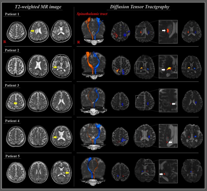
Figure 2.
T2-weighted MRI and diffusion tensor tractography of patients with CPSP following cerebral infarction. T2-weighted MR images of five patients with cerebral infarction (yellow arrows). Diffusion tensor tractography of five patients at 11 days on average after stroke onset; all the reconstructed spinothalamic tracts in the affected hemisphere originated from the posterolateral medulla and terminated at the primary somatosensory cortex through adjacent part of the infarct (white arrows) and narrowing in three patients (patients 3, 4, and 5) [reprinted with permission from Jang et al. (30)].

