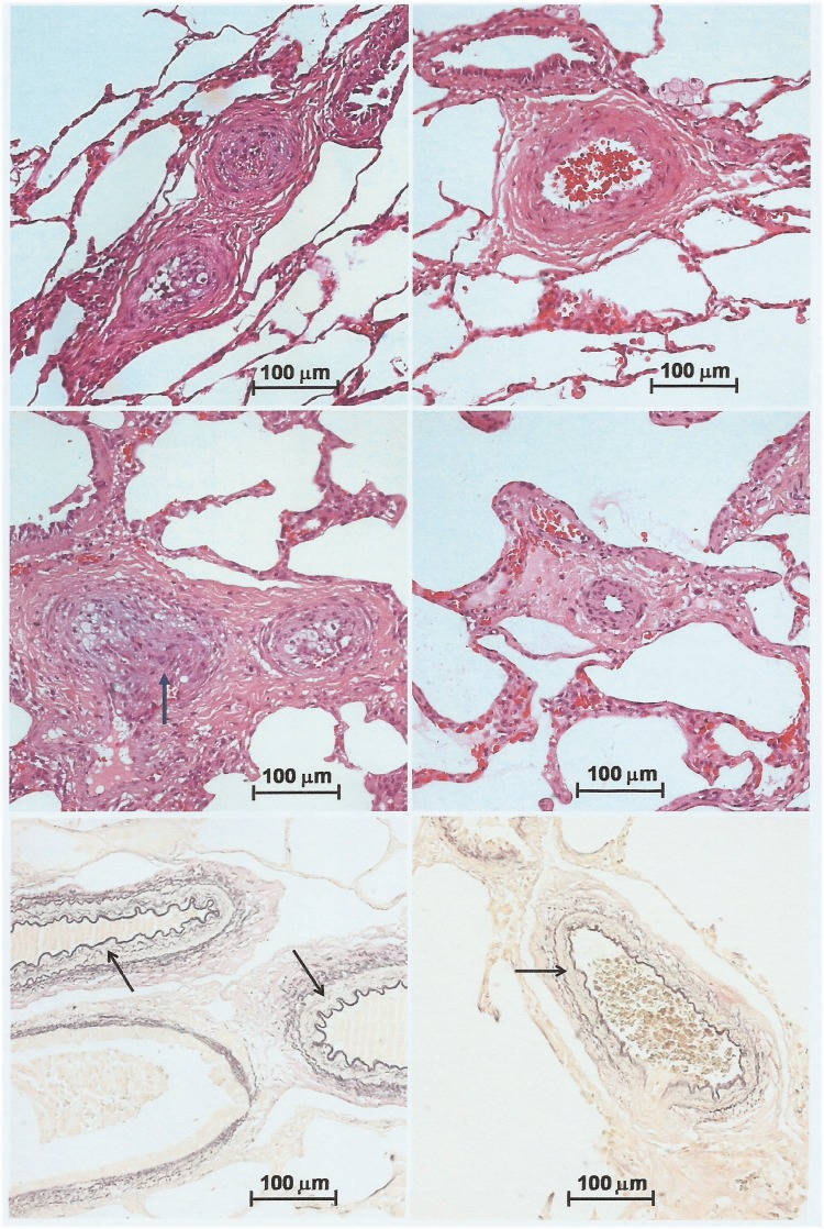
Fig. 5.
Severe and mild/moderate pulmonary arterial lesions coexisting in the same patient, thus explaining hemodynamic improvement after surgery. Top panels, 18-month-old patient with a preoperative and six-month postoperative PVR of 8.7 and 4.2 Wood units × m2, respectively. Upper left, intra-acinar arterioles showing severe occlusive intimal lesions (grade III). Upper right, preacinar artery showing moderate hypertrophy of the medial layer, with no intimal proliferation (grade I). Middle panels, 11-month-old patient with a preoperative and six-month postoperative PVR of 5.2 and 2.8 U × m2, respectively. Middle left, two preacinar arteries with intimal lesions, the bigger one showing a nodular proliferation of spindle cells, compatible with an initial plexiform lesion (grade IV, arrow). Middle right, intra-acinar arteriole with mildly occlusive intimal lesion and moderate medial hypertrophy (grade II). In upper and middle panels, H&E stain. Lower panels, Miller’s elastic stain used for quantifying medial hypertrophy of pulmonary arteries (arrows). Left and right, patients with wall thickness of arteries accompanying the terminal bronchioli of 8.1 and 7.5 (Z score), respectively. Both had normal PVR six months postoperatively (1.8 and 2.4 U × m2, respectively). In all panels, objective magnification 20×.

