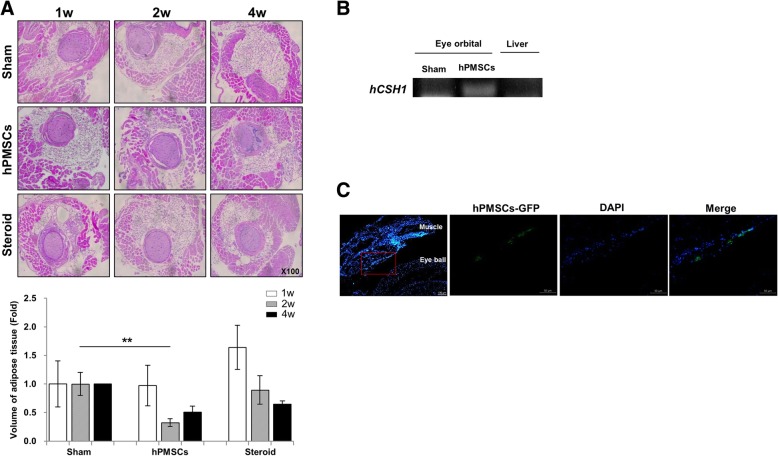
Fig. 2.
Histologic analysis of animals undergoing experimental GO treated with hPMSC or steroids. a H&E-stained section from GO mice with an expansion of retrobulbar adipose tissue around the optic nerve (× 100). Using b cDNA from mice orbital tissues and liver and c OCT-embedded mice tissues, the presence of hPMSCs was found between the extraocular muscle and eyeball. Data was presented as the fold changes (means ± SEM) of adipose volume compared with the sham of each group. Significantly different values between the groups are indicated with asterisks (**p < 0.01)

