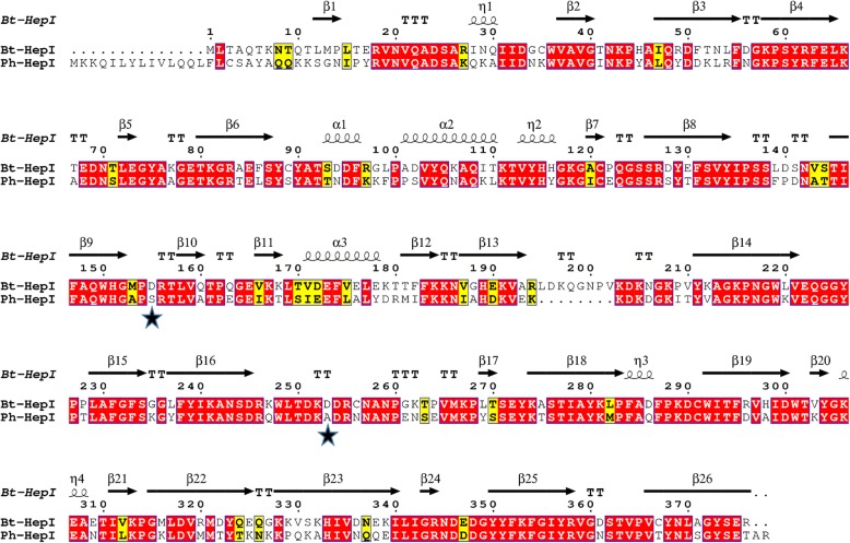
Fig. 1.
Structural sequence alignment of Bt-HepI and Ph-HepI. Residues forming the secondary structures are highlighted in the Bt-HepI sequence. Identical and similar amino acid residues are shown by white letters on a red background and black letters on a yellow background, respectively. Two different amino acids that are not conserved in Ph-HepI and Bt-HepI are indicated by black stars

