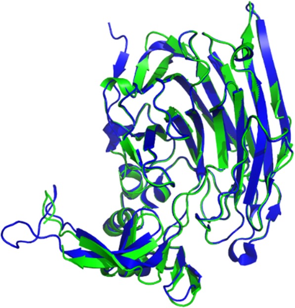
Fig. 2.

The overall structure of Ph-HepI. Structure comparison of Ph-HepI (green) is superimposed with the template from Bacteroides thetaiotaomicron (blue; PDB ID: 3ikw)

The overall structure of Ph-HepI. Structure comparison of Ph-HepI (green) is superimposed with the template from Bacteroides thetaiotaomicron (blue; PDB ID: 3ikw)