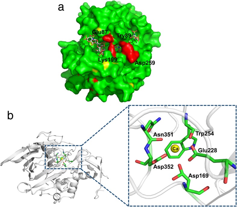
Fig. 4.

Structures of Ph-HepI bound with heparin and calcium ion. (a) The binding of heparin in the positively charged canyon of Ph-HepI-A259D is shown as a surface charge presentation. The similar cover, including four amino acids (Glu87, Tyr89, Lys199, Asp259) on the surface, is shown in red. (b) The interaction between Ph-HepI and calcium ion (Ca2+). The Ca2+ is shown as a yellow sphere. Amino acid residues Asp169, Glu228, Trp254, Asn351, and Asp352, which are essential for the interaction, are shown in sticks
