Fig. 2.
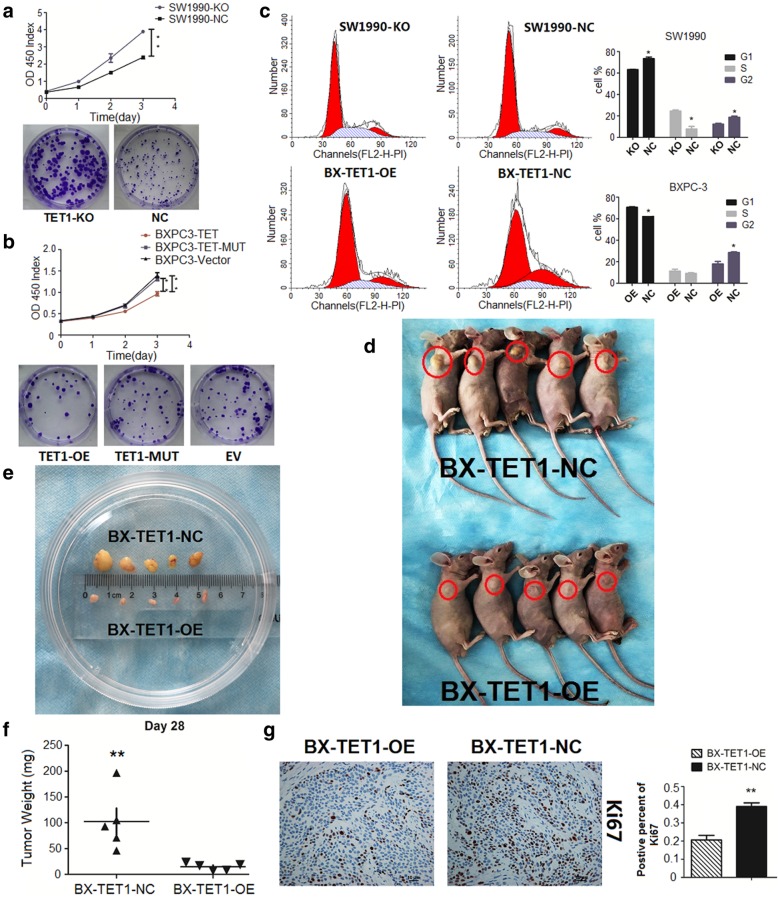
TET1 suppresses pancreatic cell proliferation in vitro and in vivo. a In SW1990-KO, TET1 knockout strongly accelerated cell proliferation (**P < 0.01, up CCK8, down colony formation assay). b TET1 overexpressed in BXPC-3 by TET1-OE transient transfection inhibited cell proliferation (P < 0.01, up CCK8, down colony formation assay) compared to TET1-MUT and EV transient transfection. c Flow cytometry analysis of the cell cycle distribution of SW1990-KO and BX-TET1-OE cells compared with their negative control cells, respectively (left). Data summary (*P < 0.05, right). d and e Subcutaneous tumor formation (red loops) of BX-TET1-OE and BX-TET1-NC cells. f Tumor weight comparison (**P < 0.01). g Ki-67 IHC analysis of tumors implanted in nude mice (mag. 200×, left). Data summary (**P < 0.01, right)

