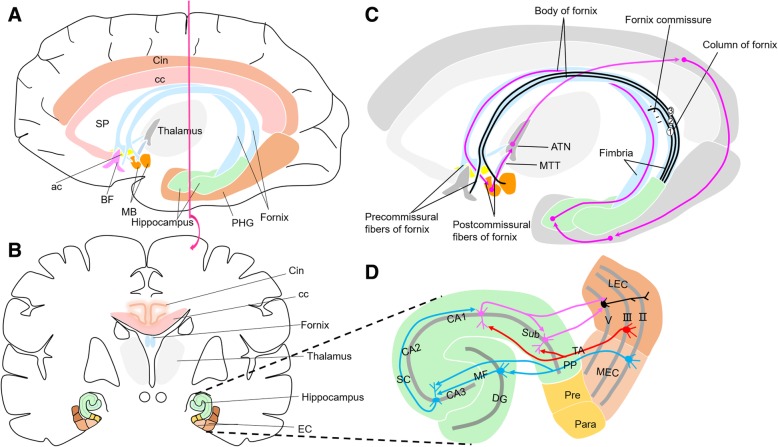
Fig. 1.
a Lateral-medial view showing location of the hippocampus and parahippocampus gyrus (PHG) in the temporal lobe. b Brain is sectioned coronally to show the hippocampus and entorhinal cortex (EC) of the medial temporal lobe. c Anatomy of the fornix and Papez circuit. Black lines show columns of the fornix. Column 1 = fibers crossing the hippocampal commissure to join the contralateral forniceal body. Column 2 = postcommissural fibers of the fornix that arise primarily from the subiculum and project to the mammillary body. Column 3 = precommissural fibers of the fornix that arise from the pyramidal cell layer of the hippocampus and project to the BF. Magenta lines indicate path of Papez circuit (hippocampus-fornix-MB-MTT-ATN-Cin-EC-hippocampus) and dots indicate integration sites. d Anatomy of EC-hippocampus network. Partial magnification of Fig. 1b with a 90° counterclockwise rotation. EC and hippocampus form a principally unidirectional network, with input from the EC. PP consists of axons from EC layer II stellate cells, projecting indirectly to the hippocampal CA1 area via Mossy fibers from the DG and Schaffer collaterals from the CA3 (red lines). TA consists of axons from EC layer III pyramidal cells, projecting directly to hippocampal CA1 (blue lines). Neurons from CA1 and subiculum (Sub), in turn, send the main hippocampal output back to the EC layer V (magenta lines), forming a loop. Abbreviations: ac, anterior commissure; ATN, anterior thalamic nuclei; BF, basal forebrain; cc, corpus callosum; Cin, cingulate gyrus; DG, dentate gyrus; EC, entorhinal cortex; LEC, lateral entorhinal cortex; MB, mammillary body; MEC, medial entorhinal cortex; MF, Mossy fibers; MTT, mammillothalamic tract; Para, parasubiculum; PHG, parahippocampal gyrus; PP, perforant path; Pre, presubiculum; Sub, subiculum; SC, Schaffer collateral; SP, septa pellucidum; TA, temporoammonic path

