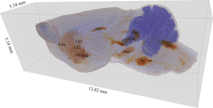
Fig. 2.
3D reconstructed image of cholinergic neuron distribution in the left hemisphere of the brain. In primates, cholinergic neurons are assigned to eight groups: Ch1 = medial septal (MS), Ch2 = vertical limb of the diagonal band of Broca (VDB), Ch3 = horizontal limb of the diagonal band of Broca (HDB), Ch4 = nucleus basalis of Meynert (NBM), Ch5 = pedunculopontine nucleus (PPN), Ch6 = laterodorsal tegmental complex (LDT), Ch7 = medial habenula (MH), Ch8 = parabigeminal nucleus (PBN). In this figure, Ch1–7 correspond to related cholinergic neuron groups in the mouse brain. Cpu = caudate putamen, Mo5 = motor trigeminal nucleus,7 N = facial motor nucleus, 10 N = dorsal motor nucleus of vagus

