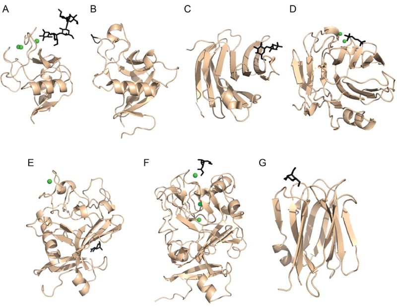
Figure 1.
Structural representation of CRDs present in human soluble lectins. A) C-type lectin domain of rat MBL (PDB:2MSB) bound to oligosaccharide ligand [23]. B) C-type lectin domain of human RegIIIα (PDB:4MTH) with residues proposed to function in peptidoglycan binding highlighted in black [28••]. C) Beta-sandwich or jelly-roll fold from human galectin-3 (PDB:3ZSJ) bound to lactose [29]. D) Jelly-roll fold of human C-reactive protein bound to phosphorylcholine (PDB: 1B09) [30] E) Fibrinogen-like domain from human L-Ficolin bound to N-acetyl-glucosamine (PDB: 2J3U) [31]. F) Intelectin domain from human intelectin-1 bound to β-Galactofuranose (PDB: 4WMY) [16••]. G) Beta-prism fold from human ZG16p bound to mannose (PDB: 3VZF) [32]. Bound calcium ions are shown as green spheres and carbohydrate ligands are depicted in black.

