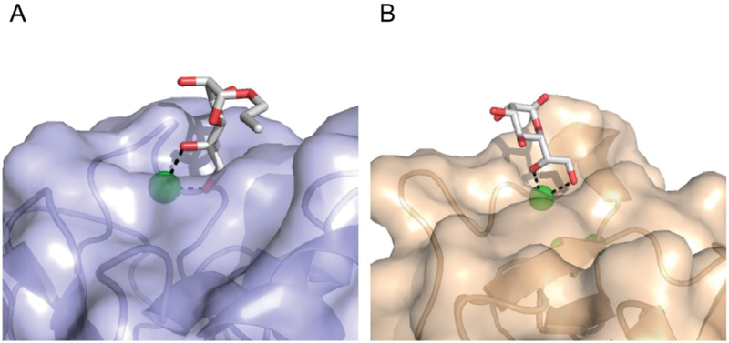
Figure 2.
Lectin recognition of exocyclic 1,2-diols. A) Surface rendering of human intelectin-1 binding the exocyclic diol of allyl-β-D-Galf (PDB: 4WMY) [16]. The aromatic box in the ligand binding site, which is constrained by tryptophan 288 and tyrosine 297, is highlighted. B) Surface rendering of human SP-D binding the exocyclic diol of L-D-heptose (PDB: 2RIB) [27]. Calcium ions (green), oxygen atoms (red), and nitrogen atoms (blue) are highlighted. The protein-bound carbohydrate ligands are depicted in the stick representation.

