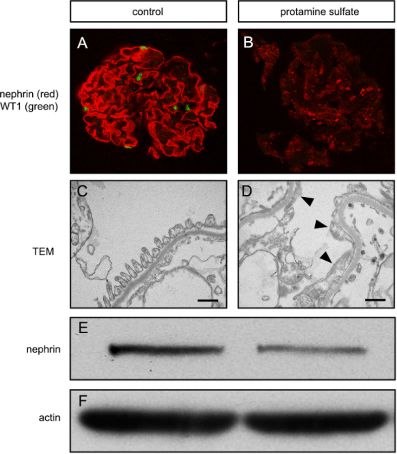
Figure 3. Podocyte injury can be induced in vitro using isolated rat glomeruli.

(A) Confocal immunofluorescence microscopy for nephrin and WT1 in an untreated glomerular culture. Note the linear nephrin staining (red) and presence of WTI-positive nuclei (green) which are typically seen when there are healthy podocytes. (B) After protamine sulfate treatment, nephrin expression is decreased and non-linear, and WT1 is absent, indicating podocyte injury. (C) Transmission electron microscpy (TEM) image showing foot processes, basement membrane, and a fenestrated endothelium typical of normal glomerular structure. Bar equals 500 nm. (D) After protamine sulfate, foot processes are elongated or effaced (arrowheads), which indicates podocyte injury. (E) Western blot for nephrin showing decreased nephrin after protamine sulfate treatment. (F) Actin loading control for western blot is shown.
