
Figure 4. Assessment of cell viability in isolated glomeruli.
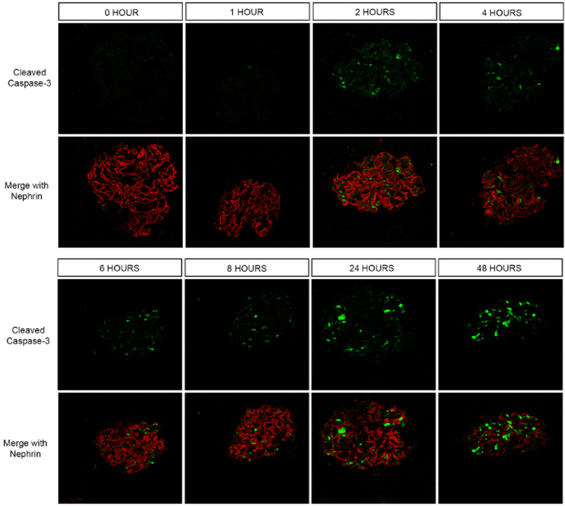
Confocal immunofluorescence microscopy for cleaved caspase-3 (green) was performed. A nephrin co-stain was performed to easily locate the glomeruli. While there was no cleaved caspase-3 positivity at 0 and 1 h after isolation of glomeruli, fluorescence signal in a few cells was noted at 2 h. Progressively more cells turned positive the longer after isolation they were examined. The greatest number of caspase-3 positive cells were noted at 24 and 48 h.