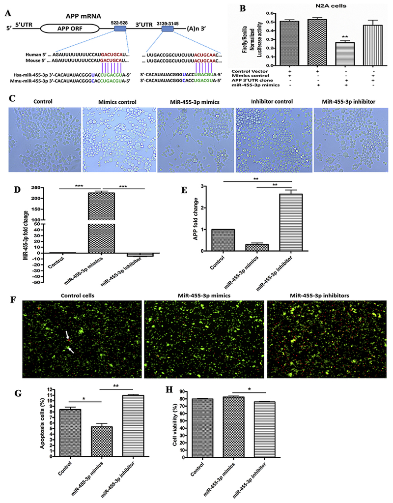
Figure 1 -. MiR-455-3p interaction with the APP gene and its effects on neuroblastoma cells survival.

(A) The two putative binding sites of miR-455-3p at the 3′-UTR regions of a wild-type APP gene in humans and mice. Region A (522-528) had 7 nucleotide binding sites, and Region B (3139-3145) had 6 nucleotide binding sites with the seed sequence of miR-455-3p in both the human and mouse wild-type APP gene (shown in green). (B) Luciferase reporter assay for miR-455-3p binding with the APP gene. Normalized luciferase activity (Firefly/Renilla) in neuroblastoma cells co-transfected with the APP 3’UTR clone (HmiT009578-MT06) and miR-455-3p mimics. Firefly/Renilla luciferase activity of the APP 3’UTR clone was significantly decrease by the miR-455-3p. (C) Representative images of neuroblastoma cells morphology after 24 hr of miR-455-3p mimics and inhibitor transfection (10× magnification). Quantitative measurement of miR-455-3p (D), and APP fold-change (E), in cells at 24 hr post mimics and inhibitor transfection. (F) Representative images of Annexin V and PI staining of cells at 24 hr post-transfection of miR-455-3p mimics and inhibitor. White arrow represents the viable (green) and dead (red) cells. Percentage (%) of apoptotic cells (G), and viable cells populations (H), after 24 hr of miR-455-3p mimics and inhibitor transfection. (*P<0.05) (**P<0.01) (***P<0.001)
