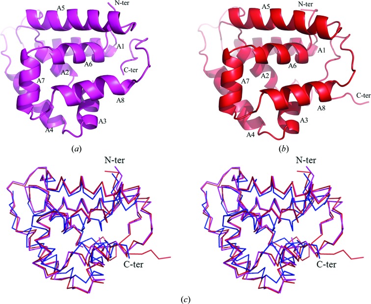
Figure 2.
The monomer structures of CaHsp104NTD and ScHsp104NTD shown as ribbon drawings. Helices A1–A8 are labeled. The N-termini and C-termini of the molecules are denoted. (a) The monomer structure of CaHsp104NTD. (b) A ribbon drawing of the ScHsp104NTD monomer. (c) Structure alignment of CaHsp104NTD, ScHsp104NTD and the E. coli ClpB NTD shown as a Cα trace in stereo mode. The CaHsp104NTD structure is shown in magenta, the ScHsp104NTD structure is shown in red and the ClpB NTD structure is shown in blue. The orientations of CaHsp104NTD and ScHsp104NTD in (c) are very similar to those in (a) and (b).

