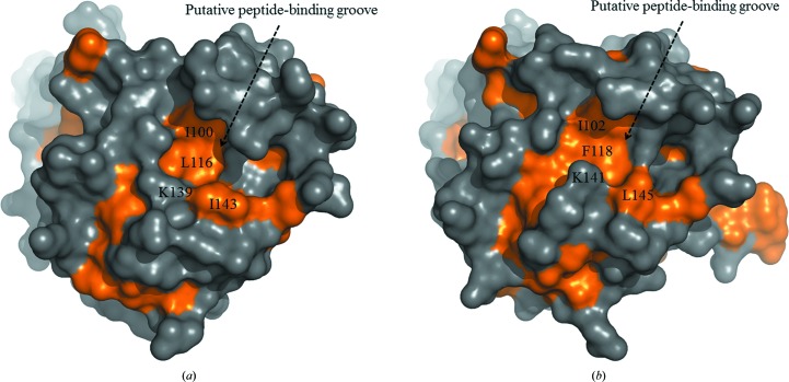
Figure 4.
The putative peptide-binding grooves of CaHsp104NTD and ScHsp104NTD shown as molecular-surface drawings. The hydrophobic regions of the molecular surface are shown in gold. (a) The putative peptide-binding groove of CaHsp104NTD is indicated. The hydrophobic side chains of residues (Ile100, Leu116, Lys139 and Ile143) which are located within the putative peptide-binding groove are labeled. (b) The putative peptide-binding groove of ScHsp104NTD.

