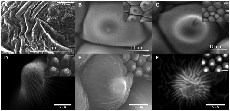
Figure 7.
Scanning electronic microscope images showing details of petal features present on the adaxial surfaces of the 6 species used for our study at various magnifications to accommodate for differences in feature size: (A) A. huegelii (6,000×), (B) S. laciniatum (3,383×), (C) L. rantonnetii (3,294×), (D) T. majus (3,406×), (E) Hibiscus heterophyllum (3,159×), and (F) P. rodneyanum (3,228×). Insets on each panel depict a less augmented version of each image. In all insets the scale bar represents 20 μm. All SEM images were acquired using a Philips XL30 SEM microscope.

