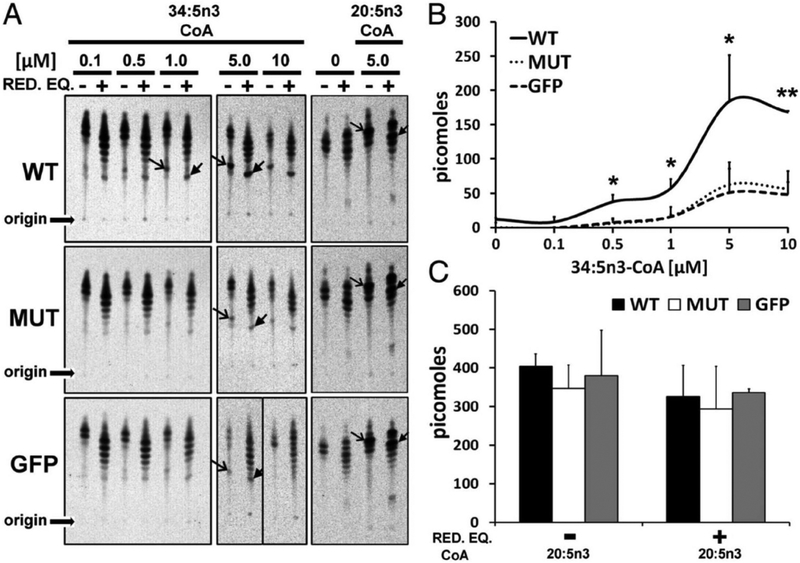
Fig. 6.
STGD3 mutant lacks innate condensation activity. (A) WT microsomes mediated the condensation of 34:5n3-CoA (open arrow heads), which was elongated in the presence (+) but not absence (−) of NADPH/NADH (RED.EQ; closed arrow heads), whereas MUT activity was comparable with GFP control. Condensation and elongation activity to 20:5n3-CoA and in the absence of exogenous substrate (lane “0“) were comparable across samples. Origin of samples spotted on TLC is indicated. (B) WT generated more condensation product with increasing amounts of substrate (34:5n3-CoA) and a maximal specific activity of 200 pmol, whereas MUT was comparable with GFP control (*P < 0.05, **P < 0.01). (C) Quantitation of condensation and elongation activities to 20:5n3-CoA shows comparable activity across samples. Reproduced with permissions from: Logan et al. (2013). PNAS. Apr. 2.110(14) 5446–5451; https://doi.org/10.1073/pnas.1217251110 © 2013 Freely available online through the PNAS open access option. https://creativecommons.org/licenses/by-nc-nd/4.0/.

