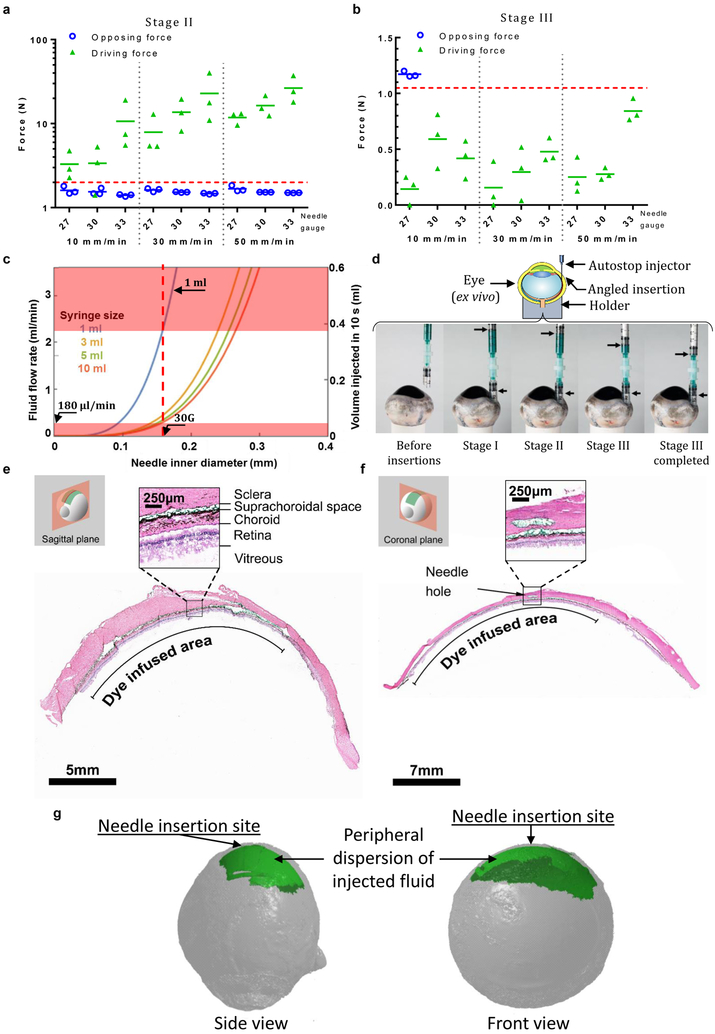
Figure 2.
The i2T2 can inject into the SCS to deliver drug to the back of the eye (a) driving and opposing forces measured in sclera for stage II (b) driving and opposing forces in stage III (c) model predicting the flow-rates that allows for automatic stop for a range of needles and syringes helps with design of i2T2 for SCS injection (d) SCS injection with the i2T2 showing the angle of insertion and position of plungers in different stages (e-f) Histology images of the injected eyes show the presence of green dye in the SCS. (g) 3D reconstructions of microCT imaged eyes following injection with contrast agent. The experiments were repeated independently (n>10) with similar results shown in (e-g).

