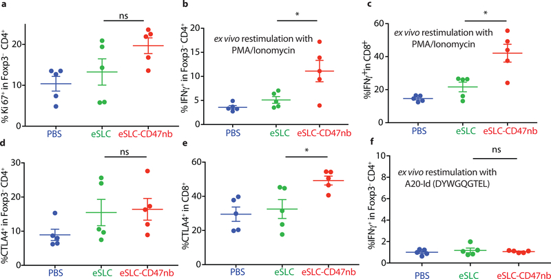
Extended Data Figure 9 |. Immunophenotyping of tumor infiltrating lymphocytes in untreated tumors following single-flank bacterial injection.
5 × 10⁶ A20 cells were implanted into the hind flanks of BALB/c mice. When tumors reached ~100 mm3 in volume (day 0), mice were treated with either PBS, eSLC or eSLC-CD47nb on day 0, 4 and 7 into a single tumor. Untreated tumors were extracted and analyzed by flow cytometry on day 8. n=5 mice per group. a, Frequency of Ki-67+ cells within Foxp3−CD4+ T cells (ns, unpaired t-test). b, c, Frequency of tumor infiltrating IFNγ + within Foxp3−CD4+ T cells and CD8+ T cells respectively following ex vivo stimulation with PMA and ionomycin in the presence of brefeldin A (*, P<0.05, unpaired t-test). d, e, Frequencies of CTLA4+ within Foxp3−CD4+ T and CD8+ T cells compartments, respectively. (* P<0.05, unpaired t-test). f, Frequency of tumor infiltrating IFNγ + within Foxp3− CD4+ T cells following ex vivo restimulation with A20-Id peptide (DYWGQGTEL) in the presence of brefeldin A (ns, unpaired t-test).

