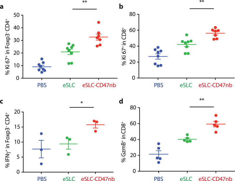
Figure 3 |. Immunotherapeutic eSLC-CD47nb bacteria prime robust adaptive antitumor immune responses.
5 × 10⁶ A20 cells were implanted into the hind flanks of BALB/c mice. When tumors reached 100–150 mm3 in volume (day 0), mice were treated with either PBS, eSLC or eSLC-CD47nb on days 0, 4 and 7. On day 8 tumors were homogenized and tumor-infiltrating lymphocytes were isolated for flow cytometric analysis on day 8. a, b Frequencies of isolated intratumoral Ki-67+ Foxp3−CD4+ and CD8+ T cells. c, Tumor infiltrating lymphocytes were stimulated following ex vivo isolation with PMA and ionomycin in the presence of brefeldin A. Frequencies of intratumoral IFNγ+ Foxp3−CD4+ T cells following stimulation. d, Percentages of intratumoral Granzyme-B positive CD8+ T cells. (n= 3–7 per group. * P<0.05, ** P<0.01, unpaired t-test, error bars represent s.e.m.). Data are pooled from two independent experimental replicates.

