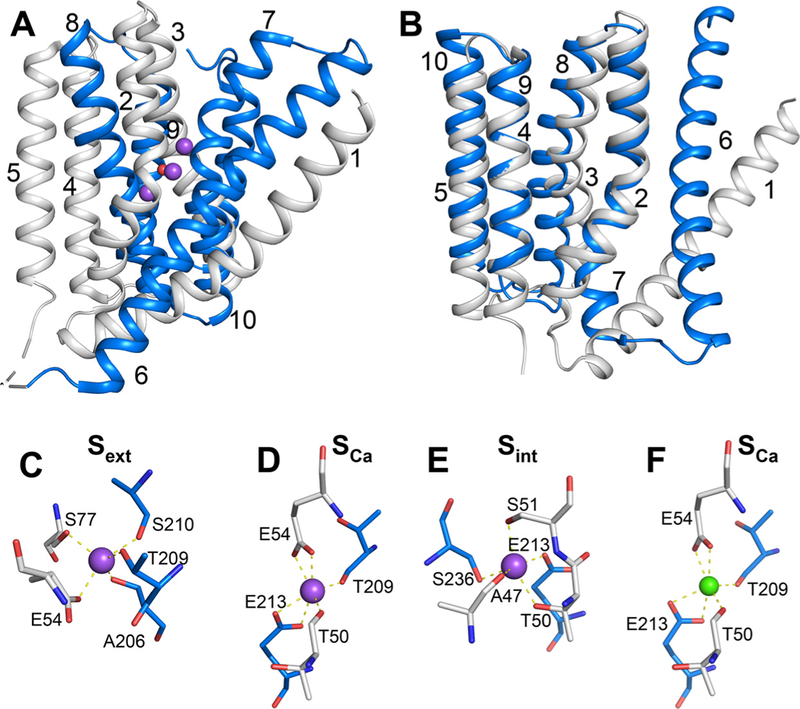
Fig. 1.

Structure of NCX_Mj. (A) Crystal structure of 3Na+-bound NCX_Mj (PDB 5HXE) in cartoon representation. The symmetry-related two halves are colored in gray (TM1–5) and blue (TM6–10), respectively. Purple and red spheres represent Na+ ions and water molecule, respectively. (B) Superposition of the symmetry-related two halves, as colored in panel A. (C–F) The ion-binding sites of NCX_Mj. The Smid site is not shown, since there is no experimental or computational evidence that this site can bind either Na+ or Ca2+. Ion-coordinating residues are shown as sticks. Purple and green spheres represent Na+ and Ca2+ ions, respectively.
